Monthly Archives: March 2018

Hydra desmosome dimensions

I found this paper on hydra desmosomes (if they can be called that, maybe better called intercellular junctions of the 3rd kind) just to compare with mammalian desmosomes. I pasted up a graphic from the diagram in this paper by Wood, in 1959, that shows the structure next to a mock up of the plasmalemma (about 4 nm thickness) and then estimated the thickness of the juxtaposed cell’s CADHERINS  ha ha… named such for lack of knowledge about what hydra intercellular adhesion molecules are — at this point –but i am looking). Interesting structure i think.  what is interesting to me is how closely the intercellular space (here about 20-30nm) matches the intercellular space of mammalian desmosomes. Another similarity of the hydra desmosomal structure is that the densities between cells are perpendicular to the direction of the plasmalemma, AND each layer of the trilaminar membrane of the plasmalemma is of a different density, this was called a plaque.

The distance between the “cadherins” is about 25nm, the intercellular distance in a non-desmosomal area of two cell connection he reports is about 20-30nm. This would be a difference with mammalian desmosomes since the intercellular space in mammalian desmosomes appears to be just a little less wide than when there is no junctional complexing going on.

I like the justification for many and for increased length of desmosomes, as this is a multicellular organism in which the need for protection from the extracellular milieu is critical.

Wood seems to suggest (which is in sync with mammalian desmosomes) that the “lamellae” as he calls them that are perpendicular to the 2 plasmalemmae of adjacent cells) are penetrated by elements of those lamella (similar to the way a transmembrane portion of a protein might behave).

Micrographs at bottom are interesting, and show the layered grid of these septate desmosomes, and while not mentioned by this author, a very close mitochondrion is present (though not touching) the desmosome in the very bottom micrograph.


Septate and comb desmosomes – who knew?

Among my favorite papers are those from the journals of microscopy and ultrastructure research way back when the TEM was first available on a commercial scale. Those guys had great eyes, nothing short of eagle vision. The limitations on figures, and words in an article were not so stringent and the micrographs and wordy descriptions abound. This included descriptions of the pretty obvious cell structures and organelles… yep, the desmosome.  The potential for examining cells was wide open, and insects, plants and fossils were favorites. The tools were primitive, yes, but the dedication was impressive. I am not too far removed from that group….I would have loved todays tools in last century’s electron microscopy boom. All that “looking” requires time, and in the warp factor 9 world of sequencing and mining….it falls behind. There is, however, no substitute for its value.

So, here is another article from the last century (LOL) showing totally different types of desmosomes (septate and comb desmosomes) the latter of which has layering with a central dense line – a little reminiscent of mammalian desmosomes (in hydra), and also desmosomes (found in silk worm), which actually have some similarities (but big differences) to mammalian desmosomes. Reference is here.
These are amazing structures, however, none of the images shown in this publication have any “look” of a connection with mitochondria as an energy and Ca+ supply, nor do they look like they have connections with the thin intermediate filaments.  In fact next to the dense plaque there is a lucent area, then there are the microtubules parallel to the plasmalemma.

One thing that these desmosomes in silkworms have is a central dense intercellular line that was not seen in insect desmosomes in the moth. (vague suggestions of a presence or absence of a central dense line (which in mammalian desmosomes is the interaction of the EC1 domains of two mirrored cadherin molecules that are part of two adjacent cells).

This article calls the picture lower right a hydra septate desmosome. I have to read more of their literature to be sure, certainly they mention the inner part of the trilaminar plasmalemma as being separated more from the outer leafelet, and the outer leafelet being justaposed by dense proteins… (in this case probably analagous to the outer dense plaque in mammalian desmosomes (i.e. the plakophilin and plakoglobin?), and I can see that there is slightly more rigidity to that portion of the plasmalemma.  Just for my own edification, i made a mash-up of some portions of one of their figures with measurements made using their own micron bar markers (red bars=50nm). Top image is a “normal” non-desmosomal membrane with membrane to membrane thickness of 18.5nm (brown bar and text) and an intercellular space between the two cells about 13nm (green bar and text). The two images below are their desmosomes. Left lower image looks like a sandwich of desmosomes (three periods=curley bracket) and red bar=50nm which appears to have three iterations (they show pictures of many more than three iterations). The micrograph (from which I have cropped just the desmosome) on the right is single, has two plasmalemma (one from justaposed cells) and a central dense line (see my line drawn in as an overlay). There are differences in the size of the intercellular space of the desmosome and the non-desmosomal plasmalemma above. Pink line is thicker than the non desmosomal juxtaposed cell membranes, extracellular space from outer leaflet of each cell is pretty similar to top image and the central dense line runs about 6 nm in.


Moth desmosomes – nice differences from mammalian desmosomes

I found this paper by Doreen Ashhurst on insect desmosomes which had some transmission electron microscopy of moth desmosomes which are clearly very different than those found in mammals. Brief notes. JCB 46: 421-425, 1970. It is a scanned pdf so the images are not that well preserved but from the text and images here is a set of general similarities and dissimilarities between moth and mammalian desmosomes (typical epithelial cell connections). The list begins at the most intracellular zone to intercellular space, beginning with the cytoplasmic structural proteins, the plaque itself (the outer dense plaque and inner dense plaque in mammals), the plasmalemma, and the central dense line where the cadherins hook up (at least there is a central dense line in mammalian desmosomes, seemingly not in moth desmosomes) .

WAX MOTH MAMMALIAN
600nm 250-300nm diam
oval shape pretty much round
cytoskeletal protein=microtubules cytoskeletal protein=intermediate filaments
MT parallel to desmosome IF parallel to desmosome
lucent area by microtubules desmoplakin molecules by IF
fuzzy outer plaque w periodicity neat tidy outer plaque w periodicity
periodicity about 20nm periodicity about 4nm apart
intercellular space about 20nm intercellular space at desmosome @10nm
desmosome has an annulus desmosome has an annulus
intercellular densities periodic intercellular central dense line


One thing seems true, the periodicities are less marked in insect (thought the X and Y configurations of what would be equivalent to the desmogleins and desmocollins are just barely seen in this photo by Ashhurst I bet they do have some relationships to mammalian desmosomes. The differning dimensions of the desmosomes between moth and mammal are really quite interesting.

Her micrograph did not have a scale bar but I used a microtubule (with a diameter of 25nm) as the standard. Red line through desmosomal outer plaque used to measure the diameter in the long dimension (presumably) of the insect desmosome. Microtubule in the upper left used to get an estimate of overall magnification of the image (microtubule at a nominal 25nm diameter). Similarly, moth to mammal, there was a slightly reduced width (height) of the intercellular space at the central part of the desmosome, than there was at the extracellular space between the adjacent cells at the points away from the desmosome. The distance within the desmosome being smaller than the distance (height or width of the intercellular space) adjacent but which is NOT part of the desmosome.  There is an annulus for the moth desmosome, just like for the mammalian desmosome.

I love that this author says…. “intermediate filaments may run parallel to the plasmalemma in mammalian desmosomes…. ” i know she saw that problem with the early interpretation…. thats fun, and she saw it way back in 1970, and the same is misjudged today.  Old habits die hard, and hardly die at all.

Classical and desmosomal cadherins: bullets

Surface proteins
Nidi for cell aggregation
Influence tissue states (layering)
Mediate selective cell-cell adhesion
Regulate embryologic cell sorting
Switches specific cadherin expression
Segregates embryological layers

Image from Farquhar and Palade below, and enlargement
It looks to me like the extracellular domains of the desmosomal cadherins (model found from some other publication obviously) fits well below the plasmalemma of the two apposed cells. This puts the transmembrane and intracellular domains of the cadherins here as a huge portion of the remaining molecule to span from the top and bottom of the punctuated extracellular space (this doesn’t include the dotted central density of the extracellular space generally called EC1) quite a ways from the plasmalemma. I am waiting to find a WHOLE molecular model of desmocollin 2 or desmoglein so i can visually “fit” those molecules THROUGH the plasmalemma on both sides of the cell-cell junction. The red circles, likely the densities at regular intervals of the connection of the intracellular portions of desmocollins/desmogleins with the plakoglobin and plakophilin?  There are some very nice tomographic images and studies of desmosomes, but they seem to fit the TEM of the extracellular molecules pretty well (not perfectly) but dont manage to show the transmembrane and intracellular portions of the cadherins with any clarity, in my opinion.  And I cant find any molecular images of the whole cadherins (all 3 domains).

The desmosome is not a “spot weld” and not a “bolt” but maybe a modified? “rivet”

A modified rivet

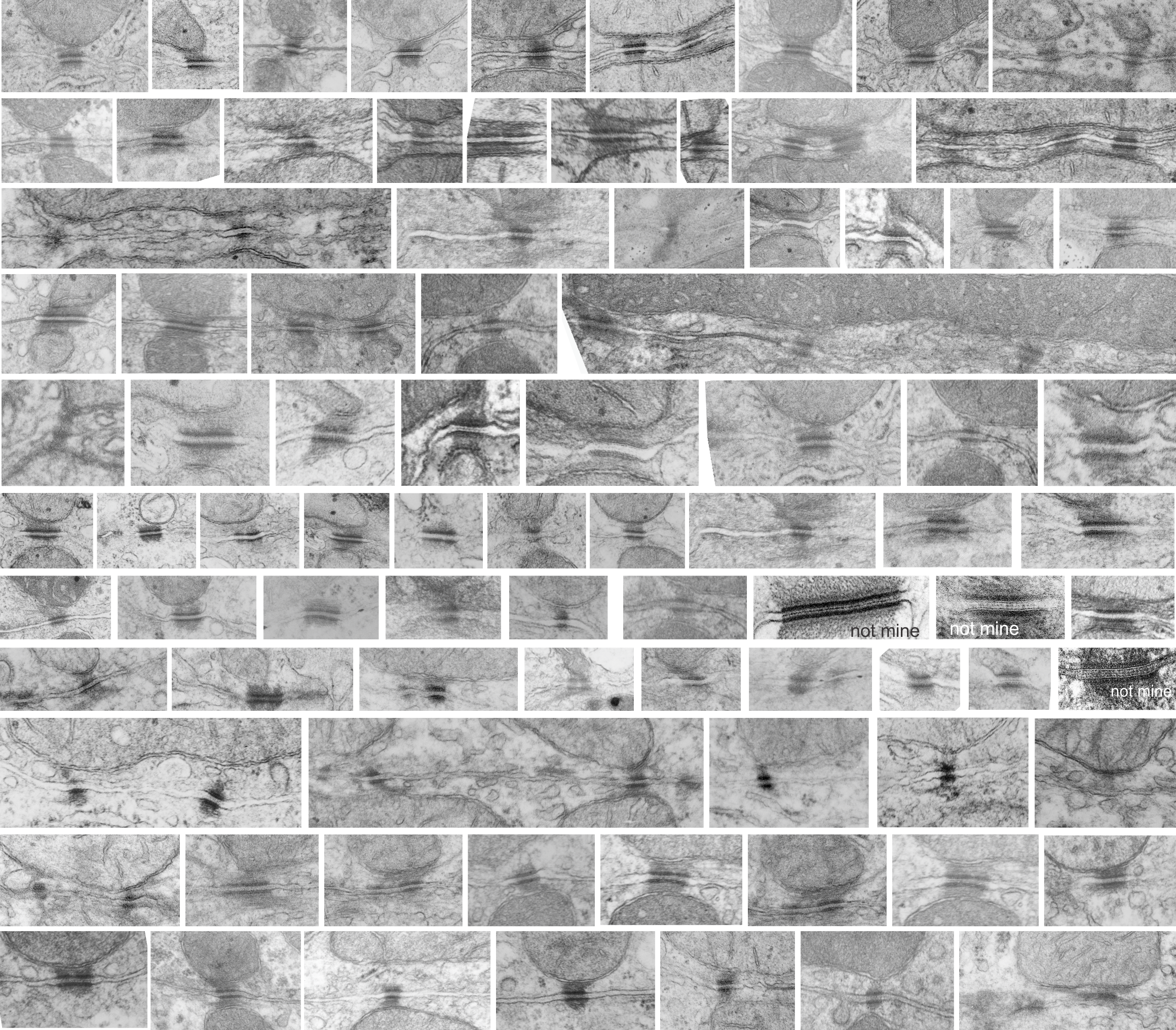
There are many names which have been given to these adhesion points, that is the desmosomes.  They occur as spots, yes, of about 200-500nm in diameter, but they are certainly not spot”welds” which would imply that the two cell membranes involved become ONE, ie, the surfaces of the two sides being joined are changed. (This is more reminiscent of the tight junction, matching the spot-weld description somewhat but totally unlike a desmosome).

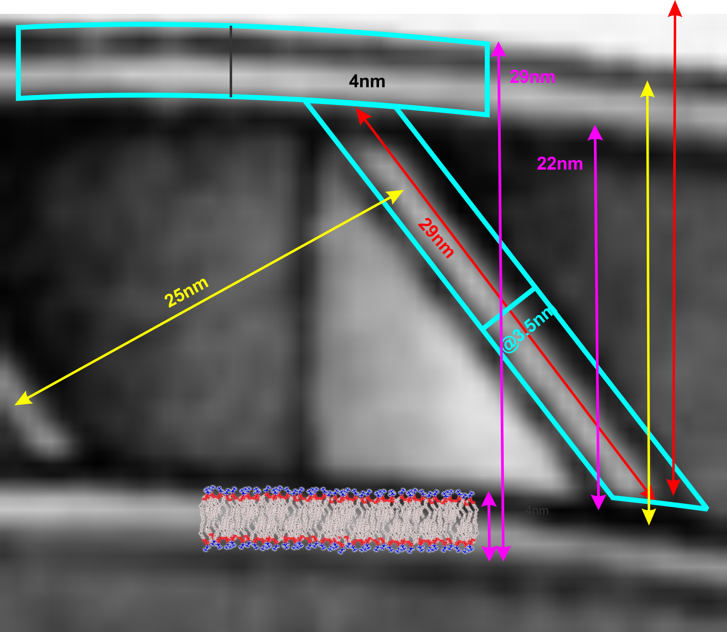
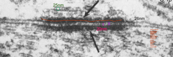
Others have called the desmosome a BOLT. This isn’t accurate either because the central line in the extracellular space of the desmosme is more like a velcro joint and not a solid inflexible mass but it is a moveable collection of various assemblies of cadherins – mainly desmocollins and desmogleins) where the center portion of the juncture (where the cadherins are bound in a central dense line with a distinct periodicity (something around 6-8nm) can be made and broken depending on ion concentration and cell signaling.  This is not at all like a solid bolt-like structure, instead it is more like a zipper.  Presumably the bolt analogy which referrers to a something “imagined” but not seen with electron microscopy was just a careless comment since a desmosome penetrating both sides of two adjacent cells is certainly NOT SOLID.  A bolt is not only solid but is temporary (or can be temporary) this on the other hand is more like a desmosome.

The desmosome is made up of hundreds of molecules positioned parallel and perpendicular to the plasmamembrane, allowing for flexibility and resistance to various stresses: shear, cleavage, tensile and peel, compressive and torsion. I am sure there are others. The desmosome might be a partial solution to shear, but it would be single shear, since there are only two surfaces involved, one from each adjacent cell.

and ensuring the  adhesive molecules penetrate the proximate side of the adjacent plasmalemma of both nearby cells. The bolt analogy doesn’t work since the central joint of the desmosome is easily “made” and “broken” depending upon cell signaling, stage of cell cycle, calcium concentration and sensibility of the cadherin molecules, ie. whether the desmosome is going to be removed or substituted, or unzipped.

  1. semi-permanent
  2. cylindrical shank  with “head” within each of the two adjacent cells
  3. similar to a buck at the intercellular junction between the cadherins from each of the two cells.
  4. major differences is that the “buck” is two sided, like the velcro…. and the integrity of the attachment sis maintained by calcium concentration (and likely many other as yet unknown factors).

A rivet however is defined as a “permanent fastener” so here we have a problem right away…. desmosomes are made and unmade on a regular basis, moved from here and reassembled there to accomodate changes in, activity, stage of the cell cycle, size, forces, maturity of the cells, and countless others.  In fact all junctions have to be totally responsive to intra and extracellular events.

Is a desmosome a little like a pop nut, or hollow wall hanger, where the is flexibility in the tethering of cadherins…. just a little, and also in tethering the intermediate filaments by desmoplakin?

Or is the desmosome is more like a bilateral (two headed) unzippable ductile lap rivet.

One of the unique features of attaching two cells with a desmosome that has no parallel in bringing two pieces together in a rivet, is that both sides in the rivet process need to be accessed….which in building….can present a problem. However, intracellular activity makes this access routine and the intercellular components are managed from the inside of each of the respective cells.

I have thought for a while about the arrangement of molecules, and its very nice parallel+perpendicular+parallel+perpendicular line up and felt that this was certainly better than any man-made adhesion rivets…. I wondered whether such a complex set of parts could actually be developed into a real live-functional construction friendly rivet.

Brazing or soldering joint….not applicable in my opinion.

Types of joints….. the desmosome is NOT a butt joint, clearly a lap joint. The spacing of desmosomes just like rivets, will be dependent upon many biological parameters, including states of differentiation.

Tell me why —

Tell me why?

If the human brain is not fully developed until age of 25 (give or take a year)(that is, it is not developed enough to have full executive, or full cognitive restraint functions)  WHY CAN CHILDREN OF LESS THAN 14 be allowed to have long guns, and any kind of gun in most states by 18.  This makes no sense to me.
I found this fantastic site. called GunPolicy.org which is run by the University of Sidney, and deals with international data on gun deaths. The graphs below I just did quickly to compare where I was born, with where I live now. You can see that the US has a big problem, but not as big as south of the border, but way in excess of the western european countries. The takeaway message can be many things.

-americans mature late in life  (haha…maybe they never develop mature frontal lobe function

-we need to change gun policy to fit american’s brains… that means, treat kids like they are really just that…. kids,

-we need to do some statistical analysis of WHO does the murdering….. young, adult, old, mentally unstable, or deviant…  and change the laws to match