Monthly Archives: June 2018

Enhanced flavoring in chicken: Simple Truth or Fake News

Here is a blog post that I like. I assume she has done her homework, I came to the same conclusions, but called Kroger to ask what their “natural flavors” meant because I suspected that the “natural flavors” included MSG or some hydrolysed veggie protein that keeps me awake at night.  They told me they did not know, and could not find out so I stopped buying chicken with “natural flavors – up to 15% ha ha enhanced”.

It amazes me how the food industry mimics our current political system which has the tag line “I can do it, i don’t care whether it is good for you or not” .   Anyway, if you eat chicken, read this.

THe blog is written by Angela Pifer – Functional Medicine Nutritionist, MS, LCN, CN, FMN

Angela is one of the nation’s foremost functional medicine nutrition and health experts, and an accomplished speaker and radio personality. Her 25 years in the health and fitness industry and the past 12 years as a Functional Medicine Nutritionist focusing in the areas of digestive health, functional gut disorders, thyroid, autoimmune and SIBO (small intestine bacterial overgrowth), have earned Angela, recognition as the go-to gut expert who can show even the most health challenged, how to restore their gut health and vitality (according to her — ).

And from anothe whistle blower (class action suit against kroger for telling the Simple Lie instead of the Simple Truth, is this blog,  also, so similar to the political climate and the spouts of insane misinformation from the current administration, not the latest of which misquotes the New Testament ha ha, people better wake up. The Simple Truth may be Fake News.

Kroger frozen blueberries: Private selection

Last week I took back a large bag of private selection blueberries. When I opened to use them it was clear by their color they were not usable (they were not purple but blue — thus the antioxidants were spent which meant to me that the bag had been left somewhere in the store for a protracted period of time and then sent back to the freezer for me to purchase (haha)).

So I saved the whole bag, called Kroger the next morning, wrote down the persons name and time and date I called and the reason I called down on my recepit. So this week I went to exchange the bag which Kroger did graciously, but i forgot to inspect the bag before I put it in my cart….silly me, especially since I have called Kroger customer service many times about faulty packaging. So here goes again, I went to open the bag, and sure enough, at the top, there was a dime size hole in the packaging.

I should have taken the bag back and raised holy moley but i did not. Here, for anyone watching their produce from Kroger, are the photos of the hole, the UPC, and the expiration date and lot.
repeated faulty packaging of Kroger frozen blueberries

More discussion on reading my slides: along with a big dose of gender and age bias from the pathologist

I was thinking about the encounter with the derm pathologist at UC last night, and while i would love to mention the name expressly, I am using better judgement and not doing that. There is something about the MD personality that is incomprehensible to me… i was married to an MD (surgeon) for 15 years, I have worked with MDs in some capacity or other for 50 years and of course what I am about to say is a generality. Thinking in generalities is problematic and it allows for bias to creep into thinking, therefore when you read the following, you must know that I am including “SELF” in that group.
Bias exists in science, as it does in all disciplines (and some lack-of-disciplines as well). In the field of microscopy as it began, there was no methodology for quantification and everything one reported in terms of histology was bias: It was biased upon previous readings, on psychological emotional and physical issues present in the slide reader at the time the slides were read, how well the slides were/are prepared, the pressure to read more and more slides faster and faster, and the biggest threat to rational thinking, lack of knowledge. So these are only a handful of really important things that influence how a slides is interpreted. The list is so long that I am not even going to give it the time.
When I began doing microscopy in the late 1960s and early 70s there were new ways to quantify cells, tissues, organelles, etc and I have used, reused, reinvented and recommended those techniques to everyone that I have collaborated with. Help from statistics programs meant that histology could be correlated with other physiologic and biochemical and behavioral measures. That said, not everyone things that morphometry (as it is called) is required for reading slides.
Not everyone even thinks that viewing slides at 100x oil is required for making diagnoses. While I sat at the student scope across from derm pathologist _blabla_ the magnification went to 60x at most, and i think 40x was probably where the objective stayed most of the time…maybe less. That for me is like hitting the tissue with a hammer when tweezers would have been the tool of choice. I asked the question, how do you see mast cells, langerhans cells, basophils, eosinophils, he said “you cant” of course i agree, but flipping the objective to 100 and adding oil would have gone a great way in making those cells visible. Not one question i asked was responded to with any intellectual satisfaction for me, for every suggestion he made i could have quoted the online general source for the same (or better) info. I expected something much more academic… ha ha… i did not expect the brush-off, so clearly signaled, nor the hostility which was directly verbalized to me with the phrase… “for the number of people you contacted” a direct reference to the two months and 10 phone calls required to get a view of MY slides. To end the meeting he put up the slides from a tray from another patient and said i guess you shouldn’t look at these… with that i said… “it is clear you want me to leave” What a bad experience. THis old man, near or in retirement, displayed a shameful bias (you know a generality) to women in science and particularly to me (also retired) that was just ludicrous.
The whole experience in Derm at UC was just not that great….maybe it is all gender and age related bias… ? I expected to be treated like a peer (which i am to him) but instead was treated like a woman whose job it was to wash dishes. How sad. UC has displayed gender bias ever since I was a graduate student…. so in 1968… to 2018, I am not seeing much of the change.

Lichen planus vs Lichenoid drug-induced lesions

Here is an article which makes one think that not all diagnoses of lichen planus are what they are recorded to be. Case in point is this report which does a host of immunological testing to find subtle differences (which would not be distinguishable by low mag microscopy and routine H&E pathology).

Just in case your thoughts go where mine do…. if a long list of drugs can cause lichen planus-like pathology, then that speaks to an environmental factor on many cases of ?? dare i say ??? mis-diagnosed lichen planus. Why not add to that a long list of environmental factors in a very similar disease a list of chemicals, such as cements, mortars, hair dyes, grouts, resins and other occupational factors that are increase one’s risk for developing lichen planus. Makes no sense why there is a line drawn between what we are prescribed by MDs, or exposed to in occupations. Just thinking here.

1) Whats more, in the cases this woman looked at, at least 25% were drug-induced… this is a huge percent which no one would argue as insignificant.
2) Orthokeratosis is hyperkeratosis without parakeratosis i.e. no nucleus is seen in the upper cells of the epidermis (the latter being typical).

Pathology report: lichen planus

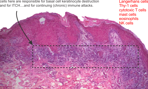
Well, that was slightly disappointing. Looking for, at most 6 minutes, through three slides was less than uplifting. The classic look: mild hyperkeratosis (thickening of the stratum corneum, or that uppermost layer of skin cells (which are without nuclei) and usually includes some additional keratin; infiltrate and apoptotic basal keratincytes; and acantholysis (the loss of intercellular connections, such as desmosomes and adhesion molecules (that can cause separation of keratinocytes in the middle and basal layers, and also signal cells to undergo apoptosis) and lots of cell debris and apoptotic bodies in the basal layer of the skin. With this there are numerous infiltrating lymphocytes (which the literature calls Th-1 cells) that apparently are able to send self destruct messages to the basal keratincytes. All this was surely evident in the slides (3 separate biopsies) from my own case of lichen planus. That said…. you ask, why are you disappointed?

Disappointed in the lack of “interest” in the real histology..like actually viewing at a magnification worth looking at… ha ha…. for an electron microscopist to be satisfied looking at 10, 40 or 60X H&E is like asking the driver of a Koenigsegg Agera RS to be happy on a scooter. While I recognize acutely (bad pun for a chronic dermatosis) how difficult it is to get through a day of reading slides I just didn’t get the feeling of thoroughness from this man, and also got a definite thumbs down when I suggested that there might be an environmental component. What gives? do the old MDs think that all of life grew up in a space-vacuum… the environment is critical.

Another question that I asked and was surprised at the answer was whether there was a good database for lichen planus cases…. he asked “database” So i asked again, to see if there were a lot of cases….so apparently there are not.

Online publications seem to suggest that lichen planus (i assume they include all variations) is 1-5% of the population? To me… that would be a lot.
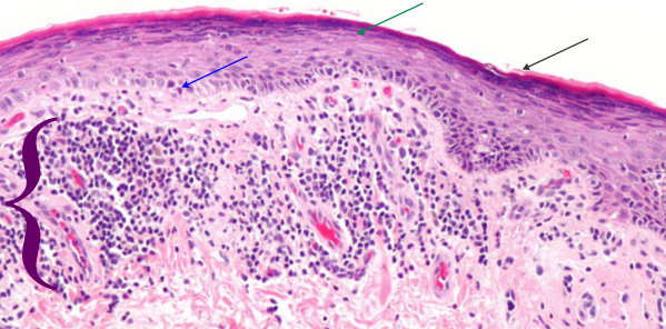
Here is a picture (wikimedia commons) which is a lot like (not exactly) what my slides showed.  Blue arrow points to keratinocytes which are undergoing apoptosis (sometimes by the old guys called Civatte bodies), green-blue arrow points to area of stratum corneum, and black arrow to the keratin layer.  White space is separation of intercellular junctions and are part of the disease process. Purple bracket = the band of infiltrating cells (for all practical purposes at this low magnification… one has to just call them mononuclear cells (which would include most cell types… ha ha). Bright pink rounded areas in dermis are blood vessels.
When i asked about langerhans cells, Thy-1 cells, melanocytes, that just drew a “no comment look” for obvious reasons, the magnification was too low, as was the interest in finding something out about these participating cells and the extent to which they might be found in different numbers in cases with different origins.  While I dont discredit the authenticity of the diagnosis from the pathologist i met with today, I do feel that the barest minimum of reporting was made, with great emphasis on “i need to move on to read other slides” and “dont bother me lady”.   This is part and parcel for being an old lady in science. It is ok to be an old male in science…. but the gender bias is alive and well.

THere was also no interest expressed in doing anything in the way of a study about the variety of immune cells, their possible connection with cumulative, and concurrent environmental triggers.  Ok, that makes me pretty energized to figure this out some other way…the pathologists here are not going to be of much help.

I have three new ideas:

I) the disruption of adhesion molecules (that would be dozens of molecules which comprise portions of the desmosomal, adherens and tight junctions, of which many participate in cell signaling i.e. apoptosis.  The adjesion molecules may be a direct or indirect response so that I will have to google.

2) The apoptotic cells: are these self induced apoptoses due to loss of adhesion molecules, or are these apoptoses  achieved through the infiltration of Th-1 cells (cytotoxic T-lymphocytes (CD8+) and NK (“educated”by langerhans cells)  which have as their goal to annihilate basal keratinocytes. A few ways to kill basal keratinocytes –by the release of perforin and granzyme or by the Fas/FasL system.

3) Are mast cells and eosinophils involved, particularly in the release of vasoactive substances, upon touching, sheering or stretching the lesions, which then triggers enormous itching.

4) another question would be, if this is indeed a “educated” set of Th-1 and NK cells, how long would one suppose that the “commitment” of those cells last.  Would it be a life long immunity gained, or something that passes quickly.

Eosinophils in lichenoid drug-induced eruption vs lichen planus

Good information to know in the differential diagnosis between lichen planus and lichenoid drug-induced eruption is the % of eosinophils.  That would be interesting. I guess I have to assume that no one in the derm path here at UC is going to make any eosinophil counts on my three slides.. ha ha. I would have to say, with H&E sections, eosinophils can stand out as cells with bright granules. I think mr. derm path that i looked at slides with yesterday would have had to up the magnification to see if this were so.  I am interested because it would increase the likelihood that my diagnosis was cement, concrete, mortar, grout related.  Publication with that data linked to yesterdays post and to this article here.
For me this is exciting information since differential counts of my peripheral blood smears has indicated an elevated eosinophil count (meaning various things, including a susceptibility to allergies) since I can remember… (so a long long time ago). This would make the diagnosis of my lichen planus more like lichenoid drug-induced (also called lichenoid interface dermatitis) for several reasons:
1) most serious lesions on shins – lower extremities, and now coming on the backs of my hands (abrasion from the leashes of two dogs when we walk, ha ha)
2) WHAT NEEDS TO BE EXAMINED in my slides…. the presence of 8-10 eosinophils per 20x field in the regions where lymphocytes are most dense. Gee… you would think that derm path could do that in about 10 minutes…. should i ask, or should i wilt back into oblivion because there is gender bias among scientists and mr derm path examiner himself would not be able to accept the challenge.

Anyway…. the point here is to make some connection between the lichen planus drug-induced (which in my case could be cement, concrete, thinset, grout-induced) dermatitis. Just thinking outloud here.

Review article: lichen planus

This article looks to be a reasonably comprehensive assessment of a disease that is varied and under-reported and annoying (i know from experience). Establishing facts and organizing data is about all we can do.
My goal is to try to figure out what the mechanism is so that I can contribute to an appropriate treatment option (selfishly for myself but hopefully benefiting others). An update on the food sorting and avoidance, I figured that after three or four weeks of not eating some foods, that if i did not see an improvement, yet even saw increase numbers of lesions that “food” was the least of my worries. But, that said, I am certainly increasing by volumes the amount of vitamin A that i am ingesting through natural sources…which leads me to recommend sweet potatoes, and red bell peppers, some dairy, since these foods have reasonable amounts of vitamin A. I am not inclined to eat organ meat, i could try fish oil.

Here is a tiny tidbit of data: When i had three biopsies (shoulder, leg, and dorsal forearm, a bandaid was put on each, and while NO lichen planus lesions grew in the biopsies…haha… they did appear under the tape marks of the bandaid. Now when Dr. Blabla says there is NO ENVIRONMENTAL component… they are just wrong.

That lesions are bilateral, mmm possibly, in my case it seems right side shins are worse than left… but so were the initial lesions created by my lawnmower (those pesky rocks, sticks, and shards of acorn) thrown back into my shins — the origin of the large lesions, and the only place that I have lesions larger than about 7-8mm diameter. Half a dozen of those on my shins are 20-30mm in diameter and 5-7mm thick. I think it is hypertrophic vesiculobullous lichen planus… two in one — LOL. I can confirm that the worst lesions are on “sun-exposed” areas of legs and arms.

This review (linked) above unfortunately does not do a good job of explaining all the nuances of the pathological architecture, structure, or ultrastructure and parrots what has been published in countless other places… though i suppose this is due in part not having a truly committed pathologist. I have tried but have not been successful (even though promised by the MD) to have access to the slides to read them myself.
When i read the slides i will take careful count of the mast cells, eosinophils and basal keratinocyte destruction to figure out if this (my case) is a lichenoid drug/chemical (in my case would be chemical) reaction rather than classical cutaneous lichen planus.
In retrospect, those who name the disease for the look of “lilchen” on the barks of trees sort of did the whole deal a disservice. And while they did not know the cause, and the cause is not known today, might have been smart not to name the disease after something that grows on tree bark. ha ha

The list of drugs which cause lichen planus like lesions is long and the number of drugs on the list given in this report would include something taken by almost everyone, and includes simple things like ibuprophen. good grief.
One thing that keeps coming to mind is perhaps a mixed-cause… that underlying condition possibly left over from a shingles outbreak, or a viral infection, or maybe even a series of bug bites that stimulate some immune response that is triggered to go into overdrive by an environmental exposure (as in my case, perhaps related to mortar, cement and ground exposure over a 10 year period). Would that someone in this insitution (there are those, like the Berensteins here in the department that are in tune to environmentally and occupationally triggered diseases, but they were too busy to take note. Just to belabor the point, the university of cincinnati does NOT take care of its own. One would think that being 50 years at this institution that I could call upon one of the MDs, (someone in the same department of environmental health, as i am, like the Bernsteins) and expect some kind of compassionate treatment…or to be able to email the pathologist who was supposed to have looked at the histological preps, Dr. Mutasim) but no such luck, in fact quite the opposite — how sad). NO perks for having spent 50 years doing research here.
Paper listed above does regard many metals as possible factors in LP reaction…so if the metals can trigger this in crowns and fillings, why not also metals found in other occupations, e.g. cement, mortar, grout, tile and similar applications. Still i could not get the attending MD in my case to do anything but scoff at the idea.

AND HERE — my fav line in this paper: LP is a complex disease and thus can be caused or triggered by genetic malfunction and/or environmental factors.

Where is the list?

Just experiencing info-disorganization-overload… ha ha

Trying to do a simple thing… to list and learn about the proteins in mitochondria.
This should be a simple task. I am looking for all proteins known to abide in the inner and outer mitochondrial membranes and those segregated to the cristae junction and the cristae themselves.

Everything…. Everything has an acronym, inconsistent and often just silly, nothing is synthesized into a neat package or publication.
I can accept that not all proteins are known at this time, but it is harder to accept that random bits of important information are not synthesized neatly into a package…
by kingdom
by phylum
by class
by order
by family
by genus
by species…
by tissue…
by cell type
by membrane position
by function
fun graphic a mitochondrionpseudocolor mitochondrion

More on Form follows function in mitochondrial cristae organization

Not much text with this post, but here is a highly simplified, not real detailed, summary of what some think about mitochondrial cristae morphology. This is very basic, i intend to make some really nice diagrams including the inner mitochondrial membrane proteins as represented in 3D by the protein databases. That will include the OSPHOS proteins, and the channel and transport proteins and the cristae junction proteins.  But for now…. and blatantly missing several types of cristae (triangular in particular) here is a line diagram of cristae and some associated properties. The electron micrographs on the “aerobic” side of the diagram are from various images of hepatocyte mitochondria just cut out of existing micrographs, on the anaerobic side, the mitochondria are selected from isolated mitochondrial fractions. Both are traditionally prepared TEM processes. Abbrev: IMS, inter-membrane space; white rim area. Outer and inner mitochondrial membranes=black lines. Grey interior= mitochondrial matrix. Micrographs, not all the same scale,


While i love the tomographic 3D reconstructions of portions of the cristae, it seems that this is sort ofwhat most of us rendered in our minds before those data confirmed the structures.


Temp files and CorelDRAW

Thank you to all of you that posted questions on “why CorelDRAW x5 suddenly began to work so slowly”…in fact it was working so slowly (as were some other programs) that it was totally unmanageable.  Just to open a file took about half a minute.  stained glass heart pattern with letter K

It took some searching, but from those posts I found out how to access “temp” files and saw that i had about 200,000 temp files (woa) some were very large, and with the cute little trick that a few of you posted, i.e. using %temp% in the windows window…. i deleted them all (i guess deleting temp files requires the temp file folder because the more i deleted the faster deleting them became — go figure) and now CorelDRAW x15 works just like new.

CorelDRAW — which my first version was given to me on 8 or 10 little pink floppy discs –version 3) and I was immediately hooked, and I found it more intuitive than Illustrator.

I use CorelDRAW for making stained glass patterns.

In praise of CorelDRAW i found it so forgiving of other software (those others with obviously built in protectionism) it was open and compatible (same with WordPerfect, way better than Word back in the day) with competitors. Even after 25 some years, I have not explored its potential — but think the drawing in symmetry feature in x8 would be awesome to have.  Maybe I will move up someday.

The pretty good export to pdf is what I have used in science, and in my own website business…  I am just saying “thank you” to the program and those who use it and figured out how to take care of some tasks, how to create macros, how to make keystrokes, etc and post that info online for the rest of us.