Monthly Archives: August 2018

Awesome video from transmission electron micrographs

Here is an awesome video… loved it.  I was searching to find the approximate dimensions of clathrin… to see if a vesicle in one of my own micrographs possibly had clathrin protein… the distance between little spokes radiation into the center of the vesicle was approximately 60nm +/- 5nm  and I found this really remarkable movie.  And i also commend that it is freely shown, and very colorful.

At the same time I continue to find TEMs from some dude in Germany whose research was very likely funded with public funds there but who also seems so insecure that he needs to “tag” that property (belonging to everyone in the world and very likely paid for by government funds) as his own… what a small mind he has– totally small and self indulgent.

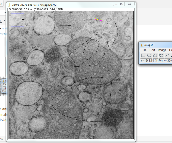
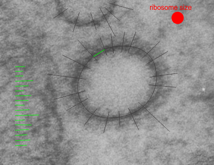
So here is the little structure I measured the proteins in….  maybe clathrin, maybe not. Measurements based on ribosome size in THIS micrograph assuming mammalian ribosome is about 27nm in diameter.   Distances between radial arms is about 60nm. Micrograph is from a 50 day wc/ii mouse. Black rays indicate location of inward projecting densities and distances between those lines at the perimeter of the vesicle were measured. A portion of a mitochondrion is on the left. Ribosome size is shown as red dot.
 

ImageJ

ImageJ is the least intuitive program i have ever encountered.  I admit to not being “wired” in the same way as many programmers, but to spend three hours and not be able to find the “select” tool says as much about me as it does about the individuals who wrote this originally and the people who added plugins.

  1. for starters, it is annoying like PyMol in that there are two or three separate visual blocks to have to click on when one wants the program to be visible… why?
  2. There is no pointer tool for select, as there is in photoshop or corelDRAW or illustrator ??? here is the set…. where is the select tool.

Why is there only ONE level of undo?  I set corelDRAW for 99 levels of undo…. and use that function to make and copy objects with slight variations all the time…. I can only find one level of undo in this program.. … what am I missing.

Pen tools…. why a dotted line as default, pen tool, why doesn’t analyze tell me the length, why cant lines be selected and deleted with the delete key or backspace?  Yes, this program may be good for some, but it a nightmare for other people.  I apologize to no one, no one can condescend or condemn… this is a very onesided interface. In my defense I have used other imaging software, and corelDRAW for 25 years – which i love,   but Image J– this program just sucks.

Using a distance and area tool ONLINE to measure inter-ribosomal distance on RER

Using a distance and area tool ONLINE to measure inter-ribosomal distance on RER was kind of interesting… There was a freebee trial of 24 hours on this one package called SketchandCalc.com which was in one sense much easier to begin with than ImageJ and L-Measure… neither of which I was able to get data from within the first hour or so. The former unfortunately does not take tif files, or pds so that is a little annoying, but it did import an 8 meg electron micrograph.
It is not intuitive in terms of setting a “denomination” for (for instance) nm, somewhere the measurement got lost? so all measures here are “relative” to my measurement of the diameter of a ribosome (well actually a mean of several ribosomes and I assumed that a mammalian ribosome was 27nm in diameter.
The whole point of making this test was to find the easiest software (shortest learning curve) to make two simple measurements, that is perimeter (and/or distance) and area from my micrographs. I am not happy with the price for the online software since this is a research project and i gain absolutely nothing from it in a personal way, it is not a business venture.

Truth be told i called a local computer repair place (been around for decades) if they might have an old Summagraphics tablet like i used back in the “day” LOL. The owner just laughed.

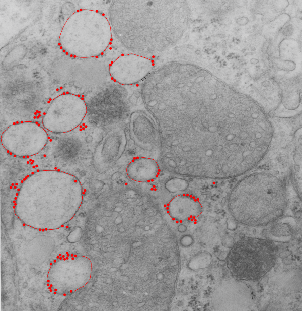
So here is the micrograph, and the measures are of the distance between the attached ribosomes… BETWEEN, because the spacing seemed in to be somewhat regular and I wondered whether there was any biological importance to that. At the same time i took the opportunity to measure several points at a mitochondrial-RER membrane close encounter.

10,000,000 in some cells x couple billion actively synthesizing cells = a whole lot of ribosomes.

Top micrograph (18406_78375_50d_wc-ii no NAC) shows dilated RER (a combo RER-SER with interestingly spaced translocase (?) attachment sites. There are other characteristics too, the length of the string of ribosomes. Red dots are ribosomes (on micrograph below); distance between attached ribosome strings is about 273.6nm +/- 27, n=30, so the SEM is 10%, that is not really that bad for biology and there might be something to this; perimeter of RER vesicles= is about 1500nm, n=10; mean area is about 1600nm2

Verge of a Dream: unorientation

Climbing up the river’s
shoulder. With spent
hours in faze. Except
for you wearing a
pattern off the shoulder.
I want to return the
unbalanced borrowings
carried by vespa til
they fall and fail.
The authorities don’t call.
So unrelenting stay
the conflicts and confusion.
I should see
the contentment in
spiritual order that
shrugs off my un-
orientation.
RLB August 03, 2018

Verge of a Dream: At the orpheum

At the orpheum
I want to today
Make the rabbit disappear.
Alone on the bus seat
The counselor observes
you are very pink today
The nurse looks at
my arm. You didn’t
use today, question mark.
I want to be on time
parking near sixth and
styx. The adviser holds
my wrists. Reporting by
form how I stole and
could do worse. I am
In the class tonight
hoping that I read
the lesson. And
at home
I can paint hexagons
orange, suspended
In a red cloud.
RLB August 01, 2018

Lycoris sqamigera: in the midst of chaos

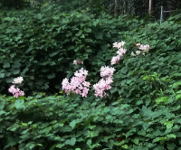
This amaryllis has to be one of the heartiest bulbs ever: here in my neighbor’s yard under the massive amounts of overgrown grape vine, poison ivy, euonymous, thistle, nettle, english ivy, and wild strawberry, the blooms appear, as a testament to order and beautify, as they stand like the only wonderfull objects amidst the three abandoned cars, stacks of tires, trash cans, inverted cement mixing tray, broken steps and falling gutters. How orderly they are, and persistent (at least 30 years), even in prospering, in the mess and bramble.


Certifiable? or does rRNA on RER rock

I was looking at liver electron micrographs, looking for mitochondria, trying to see whether cristae juntions are such a noticeable feature as is claimed, boy I can not find them easily. But in “boredom” not really boredom, more frustration, I decided to punch in a “kick” beat to the spacing of ribosomes along the RER membrane.  I hope this fun time with Fruity Loops doesn’t distract me from doing real science…. as I can see how fun it would be to use a top hat, steel drums, cymbals, for some ribosomes a little further away, and a base beat for those that are very clear and a blip for those that are faded out and then play two tracks together (opposite sides of the ER membrane being a separate track each). Now you know why I chose the title for this post “certifiable” (i am not crazy, as sheldon cooper says, my mother had me tested),

Ultrastructure of some mitochondria and RER associations

It is really likely that some of the patterns in electron micrographs (even though they were taken decades ago) can still reveal some of the unique ultrastructural architecture that cells possess, even new and useful data. (This is an apologetic of sorts, since I don’t have the latest in tomographic equipment or a wet lab but still find amazing structures in the old TEMs i have lying around.) So these images are offshoots of a previous post on mitochondrial substructure, and I noticed two new things in this same micrograph. That is:

1) that one of the mRNA and ribosomal spirals has a dense center and from it radiate spokes (red arrow in left and top middle images) and…

2) there is a portion of mitochondrial matrix? intra cristae? area which is clearly organized into repeating parallel layers with central dots.  (this is best seen in the images to the far right. The boundary between mitochondrion and RER is black dotted line, lower images on right; black box on left is area enlarged (top and bottom middle) one with contrast enhanced (bottom) and one set of mRNA+ribosomes spiral, and black line marking line likely mRNA, and to the left at the red dots a vertically oriented multilayered organization of mitochondrial (membrane?) cristae…  the black box surrounding this particular area of interest denotes the two images to the most right.  There is no question about the organization of vertical-parallel lines, and dots.  These are the blue dots in the two lower right hand figures. Vertical lines with vertical organization of the rounded densities is quite striking (upper right). This is mouse liver, neg 6118, block 5220 and from my notes it received tween and PP5 at 50mg/kg and this is a 72 hours necropsy. Right top is same as right bottom, where the central dense dots and outer dense line – substructure is observed.