Monthly Archives: November 2018

Total SP-D dodecamer measured length using arc angle dimensions

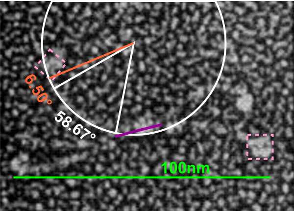
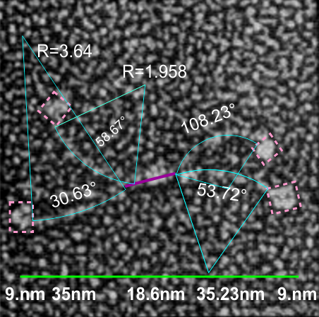
If i use the distance between two trimeric arms of SP-D seen in dodecamers as the off measured 100 nm width of the molecule, then measure the arc angle of each arm (arc angle length determined by [ 2pi R  (central angle/ 360) ]  this increases the total length (stretched as if a planimeter were used) of the separated collagen-like domain of the dodecamers at about 39nm for each half.  (n=14 individual dodecamers with n=4 arms each), or the mean of the lengths of the two sides as separate individual measures (n=58 *one dodecamer had what looked to be five arms and was included).

The mean dimensions in nm of the N terminal domain, taken as one measure probably includes the N terminal as well as some small portion of the collagen-like domain, still both tethered together tightly (that is, the N terminal and a portion of collagen-like domain) and is about 19nm. The measurement spanned the area between bifurcation of the collagen-like segments on either end of the “joined” N terminal domains. (purple line)

The CRD measures 10.8nm whether determined by the mean of the length of the two sides, sorted by dodecamer (n=14 dodecamers, each with 4 CRD), or the mean of the lengths of the two sides all as individual measures (n=58 *one dodecamer had what looked to be five arms and CRD and was included).

A total dimension of about 119nm (the individual image shows a total dimension of 112.7nm – below). The very same diagram is shown above and below, one made before i decided to check how to measure the curvature as part of the length of the trimeric arms, the other after.

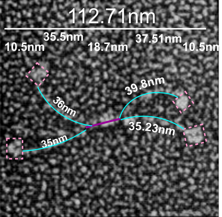
Here is the image posted earlier, where the arc length is NOT calculated, but just the straight line distance between where the trimeric arm diverges visibly from the center (Nterm plus whatever collagen-like sequences are there).  When measured in parts, the total length of the molecul below measured at 107nm slightly above the 100nm which I constructed as the diameter between two CRD lying on a the circumference of a 100nm circle.

What is pretty clear is that there are not that many, if any, accurate diagrams out there that have the proportions of CRD to thickness of the collagen like portion, or the curvature, or the length of the four segments of SP-D right.

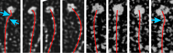
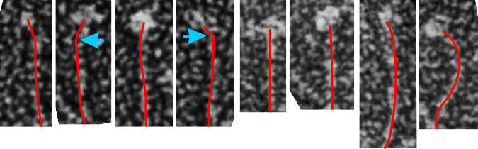
One thing for certain, is that shadowed and AFM images of SP-D show that the collagen portion is certainly NOT straight, as referred to in the chapter Alveolar Epithelium and Plumonary Surfactant, by Robert J. Mason MD and Leland G. Dobbs MD in the textbook Murray and Nadel’s Textbook of Respiratory Medicine, 8, 134-149.e5.

I could continue measuring all the dodecamer images i have collected using the arc length, central arc angle, N terminal (and whatever else) bound central segment, and size of the CRD but i don’t think adding those new data is going to change the outcome.

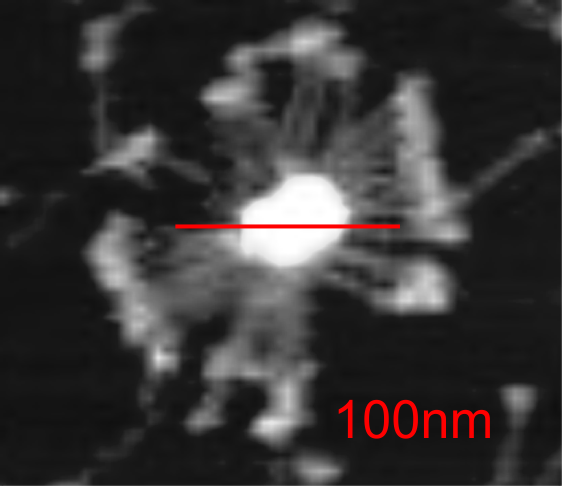
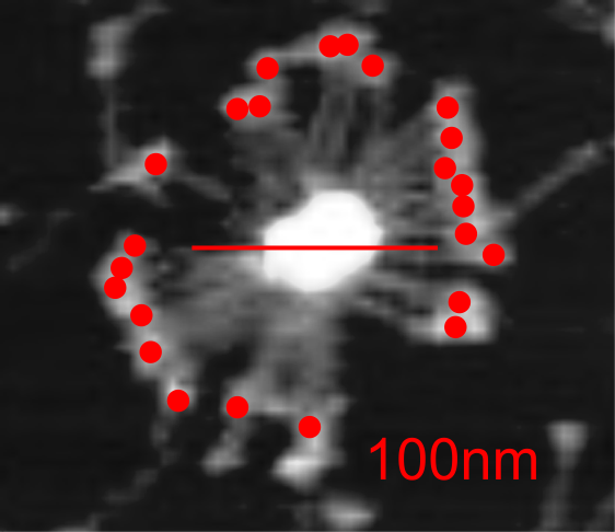
Here is a mutant surfactant protein D fuzzyball (from Kevan L Hartshorn, et al – with the mutation in the CRD so not likely affecting the overall dimensions much) which is greater than their own micron marker would designate as 100nm. This image was cut and pasted into photoshop from their figure, a dupllicate made, top layer increased in contrast, decreased in brightness, and layered at 50% transparency over their original in order to show the arms of the dodecamer and the CRD better. This is a mutant SP-D, according to them, with two substitutions in the CRD. (Recombinant human SP-D having a Met at amino acid 11 (and Ala at amino acid 160).

Counted the CRD just for the fun of it.

Arm kinks in surfactant protein D dodecamers

Surfactant protein D is a terrific molecule, and the assorted arrangements of multimers has been described somewhat but I have only found one publication which actually tries to figure out details about the unique shape of the collagen-like domain. The carbohydrate recognition domain and the coiled-coil neck section has been studied – ad nauseum and the RCSB has countless molecular models. The N-terminal has been a little less studied, even less the collagen-like domain.  I am not really sure why that is since there are dozens of images of SP-D trimers, dodecamers and multimers (fuzzyballs, fuzzy balls) from which to determine length and curvature of that domain.  At anyrate, there are some images which show kinks at the neck (a comment typically included in descriptions of SP-D, so that is not new) and at least one paper suggests there might be something going on at the glycosylation site (closer to the N terminal).  Here is a group of just for trimeric arms (cut and rearranged from dodecamers (images from manuscripts that I did NOT write – thank you for those) and it becomes clear that an assessment of angles in this kind of order might be useful. One kink, maybe two kinks (see blue arrows on left).

It is pretty clear that the length of the collagen-like segment that show up right at the crotch of the bifurcation of the arms of the dodecamer is not going to end up the exactly length of the whole collagen-like portion. It seems at this point that the there is N terminal association of the four trimers and then more close binding for a greater length up the collagen-like sequence.  The proportions of the micrographs just don’t add up (especially visually) to the dimensions given in the literature for the collagen-like sequence. In particular too, the diagrams are way off. This begs the question, where does the central ly bound portion end? and does this make a difference in the shape of the center of the fuzzyball made up of 6 or 8 dodecamers? as i mentioned weeks ago on this blog.

Measuring the arm length of SP-D dodecamers

This might work…. one molecule, using the 100 nm marker provided in countless publications (yep that is a leap of faith) and independently measuring the arc distance of each of the arms of SP-D dodecamer directly from this particular micrograph (the two CRDs as rectangles with a mean of the 16 sides total, a measure of the tethered central N terminal domains, (no neck measurement because i these images they are not really discernable). The total was close, for the first sample measured and calculated per the diagram below.  Total of the parts added is just over the presumed measurement, of 100nm.

This looks simple, but it took a while to figure out… but I can use CorelDRAW for all measurements on SP-D molecules

This looks simple, but it took a while to figure out… but I can use CorelDRAW for all measurements.  This program is amazing and I could hardly survive without it. Thanks to my son danny for introducing me to it way back when he was just a kid working at a deli that for some reason used one of the very first versions… CorelDRAW 3.  I should buy the upgrade (haha).  So measurements of molecules and structures that have been photographed with electron microscopy (or atomic force microscopy, or techniques which shadow or coat molecules to highlight them are rarely made.  Some of the really great “oldies” that formulated the basics for morphometry inspired me 40 years ago, and making what is usually a “subjective and only visual” science into something that can be graphed and analyzed has been my goal (forever it seems).  So working on the shapes of two surfactant protein molecules (SP-A and SP-D) it became clear that no one really paid much attention to how the represented these molecules graphically… some even represented (and published) them with such “license” (which certainly could NEVER be considered “artistic license” so erroneously that it was adding misinformation to other articles whose authors were using them as models.  FAKE NEWS.

It has been great fun working with images of surfactant protein D for TWO reasons:  1) the totally ridiculous diagrams that have been published… i wanted to set straight with  something more accurate and 2) these molecules can be very important in the efforts to tailor new synthetic – fullerene-type innate immune molecules to best opsonize  viruses and bacteria which enter the lung.

Previous posts show center arc angles in dodecamers, and also the angles of the collagen-like portion of the SP-D dodecamer “arms”, but other things need to be quantified.  Diagram below indicates what I think i can measure, and that is the arm length of the collagen-like portion, maybe also the length of the coiled coil neck region, the varying shape-size of the CRD, and the mean length of the N terminal.  PER THE IMAGE below.

Using the center arc angle of the collagen-like portion the formula found on this site can be used to determine the arc length. Similarly, the neck arc length.  the purple line is from the branch of the dodecamer pairs, and it seems actually to vary quite a bit.  THe CRD are easy to measure just as with width and height (since it is a trimeric structure (anticipating from the molecular models on RCSB to NOT have equal width and height, but vary in orientation sufficiently in the micrographs to present with some variation.

There are two kinks for sure, one was predicted by some researchers to be close to the N terminal, and I see one (sometimes represented in various diagrams) at the coiled-coil neck region.  I havn’t decided whether to measure that or not.  So many studies have shown that the dodecamer is about 100nm that has been used as the standard measure from which other measures are made.  EDIT:  so neck measures are not going to be possible, and will be part of the CRD measurements.

Ecclesiastes 9:11

HOW SPOOKY IS THIS…. Ecclesiastes 9:11 and 911 and to call nine-0ne-0ne.
This is what Solomon says.
I have seen something else under the sun: The race is NOT to the swift or the battle to the strong, NOR does food come to the wise or wealth to the brilliant or favor to the learned; but time and chance happen to them all.

Followed by our current political situation:
Moreover, no one knows when their hour will come: As fish are caught in a cruel net, or birds are taken in a snare, so people are trapped by evil times that fall unexpectedly upon them.