Category Archives: Ultimate order, the cell

The beauty and order of life is astounding.

Variety: the whole spectrum of lysosomes/inclusions of E2 in an alveolar macrophage

Variety: the whole spectrum of lysosomes/inclusions of E2 in this micrograph of an alveolar macrophage (at least the whole variety I have seen so far). It includes lysosomes (LE/LY/PFC) which probably could be classified all the way from golgi vesicles to late endosomes since it seems that PFCs in general are able to work their way into the whole ER system (though to date I have not seen anything that looks like a PFC droplet in any ER which has ribosomes….  still looking). There are droplets in a “hollow” meaning no lysosomal enzymes yet, on the right but still having E2 droplets with the coating they must pick up from somewhere (maybe alveolar surfactant in the alveolar space?, being lipid and E2 being slightly lipophilic), to the dense small lysosomes with many E2 droplets on the left, to a single E2 droplet on the lower left, and a dozen droplets which look like they are hanging out in the cytosol with no membrane boundaries, to bottom center structure which looks like two E2 droplets in a lysosome with a lamellar type surfactant inclusion.

There is no paucity of free ribosomes in this cropped image, and size can be inferred from the approximate 27nm diameter of a ribosome (red dot), to the larger droplet size (bar=270nm).

Countless PFC droplets in alveolar macrophage lysosomal body

The lysosomes in this alveolar macrophage from a mouse which breathed E2 for 3 hours and then allowed to recover for 5 days are awesome.  The macrophage has produced enzymes which re-emulsify the E2 into very small (in most cases) droplets within the lysosomal structure. In addition, the enzymes make a border, which is very difficult to distinguish from the trilaminar membrane which surrounds the lysosome proper.  I don’t know how to explain this look of a double membrane, but figure it is partly a physical interaction between the E2 droplet (and not unique to E2 but seen with many other perfluorochemicals) and its hydrophobicity, and slight lipophilicity.

The smallest spheres of E2 are down around 35nm diameter, and size can be compared to something just larger than a ribosome (@27nm). Picture on the top is the unretouched (i may have removed a scratch with the band-aid tool in photoshop but nothing else), and it is not too great an image (scanned from the 31/4×4 acetate negative), but interesting still, and providing lots of information. Image below is one where I have highlighted the E2 droplets in a single membrane bound lysosome and embossed them.

The dark structure (also rounded) in the upper right hand corner of the images is what I think is a phagolysosome that contains mostly phagocytosed surfactant lipids (some layering and myelin-look is seen within this structure) from the alveolar space. There are also two mitochondria near center top. It also seems likely that some of the surfactant engulfed by alveolar macrophages would find its way (during re-purposing or re-cycling) into such lysosomes containing E2 droplets.

electron micrograph alveolar macrophage E2 liquid breathing perfluorochemicalelectron micrograph alveolar macrophage E2 liquid breathing perfluorochemical PFC highlighted

Is this E2 in alveolar endothelium?

Loooooking over an old negative of lung from a mouse that breathed oxygen bubbled liquid perfluorochemical  (in this case E2, which is a freon) I spotted what could be, maybe, seems almost possible, droplets of E2 in the endothelium (alveolar sac endothelium).  I selected portions of the image out and pseudocolored them for easy identification: Red cell within the capillary lumen, red, endothelium orange, lysosome/or vacuoles that might be E2 and comparison with those which are in a nearby alveolar macrophage, green, and the macrophage cytoplasm is purple. There are some reasonably large E2 droplets which are probably unequivocal (is that not an odd combination of words?) with more distinct membrane boundaries, and absolutely NO fuzziness within the inclusions but also having the distinctive “black cap” of lysosomal enzymes off to a little blip on some side or other of the droplet.   This is NOT seen in the endothelial inclusions…. I will look for less equivocal samples but the similarities between those and additional ones found in the macrophage is clear. 1351_4840_mouse_E2 breathing for 3 hours, 48hr recovery, capillary_endothelium and two macrophages.


E2 lysosome “Looking at U”

Could not help but post this cute little lysosomal structure in a macrophage of a mouse that liquid breathed E2 (see previous posts for chemical structure) for 3 hours and was allowed to recover for 48 hours. This lysosome is a tiny portion of an electron micrograph of a macrophage in the alveolar space of the lung.

The scope of lysosomal variation in these macrophages is really quite astounding.  I see lysosomes with a tiny bit of enzyme material localized on one side of the membrane bound structure, and others, like this, with maybe 4 or so tiny perfluorocarbon droplets (which looked like two eyes to me), and massive amounts of enzyme (all that medium grey in a round membrane structure). A library of these variable organelles is what I am attempting to do at this point.  E2 is capturing my attention. If you look closely, around the periphery of the lysosome there are small circular areas, which I have no doubt are the tiniest lysosomal droplets down around the size of 20-30nm in diameter.  The “eyes” that are droplets here are much larger than that, closer to 100-200nm). Highlight in the eyes and the dark pupil i did in photoshop just to have fun, but the eyeball itself is E2 and the “nose” is enzyme density.  In the upper left corner of the image there is another lysosome with a round E2 droplet (about 75nm diameter) with some layered darker substructure.

It is possible that part of the lysosomal material here is unique to lung since alveolar macrophages (which this is) tend to uptake surfactant in large quantities, and it seems that some of this lipid material (perfluorocarbons have different lipid solubilities, and this might contribute to the mixture of electron dense (osmiophilic) nature of the lysosomes. I might call these MVE/LE/PFC, but that is too cumbersome, hence forth, just lysosome. Besides the specific proteins and enzymes and pH in these structures has not been measured in this archival material… thus there is no way to accurately classify them so I will call them lysosomes.\


Inert and partially inert particles in lysosomes

Here is something that might be close to looking at the endosome-to-lysosome pathway taken by perfluorochemical droplets in experiments either of liquid breathing or infusion of perfluorochemical-based blood substitutes.  I this particular article (Zhou et al, Int J. Nanomedicine, 2010 — might be a good journal to submit the PFC papers upcoming).  It is nice to see that the endosomes look a little like the dense, highly enzyme filled, MVB/LE and lysosomes seen in macrophages and other inclusions in cells exposed to PFC.  I am excerpting and editing one of their TEMs… without permission, but giving them credit, to make this parallel. Particles (Poly(d, l-lactide) (PLA) and poly-d, l-lactide-poly(ethylene glycol) (PELA) with PEG weight ratios of 10%, 20% 30%? I think this is all) don’t show a two phase morphology with their SEM images, but clearly with TEM there is a dense core and a lighter outer area. This is in total contrast to the “footprint” of PFC, in the image on the left which is basically “completely electron lucent”. But there are some similarities in the lysosome (called that just for simplicity) in each of the two images, though the PFC lysosomes (on the left) really are densely packed with enzymes, maybe even more than the nanoparticles (on the right). SEM of nanoparticles, see inset on the right.

Lysosomes, diagrams, hysterical typo

hysterical typo on scientific diagramI hope I am able to laugh at my mistakes, as this individual needs to laugh at their typo.  What…  ha ha… a lysosomal rapture…. no kidding,  waiting for the rapture myself, and hoping to be among those taken into the ether.  Just in case you missed it…. lysosomal rupture would be, i must presume, what was intended.

Perhaps this book chapter is source of the “rapture”

Not typical lysosomes: Do engulfed PFC droplets migrate up the ER

Looking at lysosomes in the lung of mice which have breathed perfluorochemical (PFC) liquids is interesting. These are old archived micrographs from the early 1970s but have a lot of valuable information about the reactivity of perfluorochemicals in the body (the latter being touted as totally inert.  Regardless of what science says about the reactivity of PFC they do cause a mountain of effects in humans, not the least of which is an immediate uptake by the reticuloendothelial system, massive sequestering and dumping of enzymes into the resultant lysosomes, and a kind of partitioning, or re-partitioning, or re-emulsification of the PFC, depending upon whether it was breated as a neat liquid, or infused as an emulsion with other agents accompanying it.

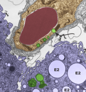
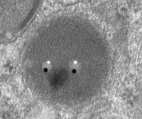
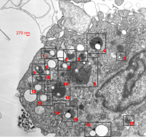
So “case in point” is this alveolar macrophage, picture taken from the lung of a mouse which breathed E2 for 3 hours and was allowed to recover for 48 hours then lungs taken for electron microscopy (and probably chemical analysis, though I have no clue where those data are at this point 40 years down the pike).  I just know that my negative number is 1351, and that block number is 4840, magnification was 5,000 (Zeiss 10) date 2 17 1975 at 60kv.  Mouse weighed 16.85g, breathed E2 for 3 hours on 2 3 1975, and was processed on 2 5 1975, which equates to 48 hour recovery. The sample of lung was taken from the R, mid lobe.

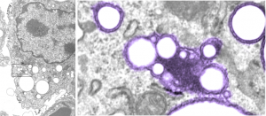
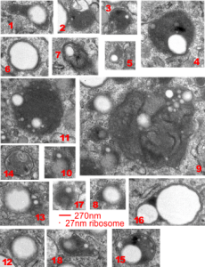
The image below (L) is a portion of the original negative (scanned as a transparency from the film) with an enlargement (box demarcates it) on the right where I have “pseudocolored” the protein densities with purple, which surround the empty footprints where the PFC was fixed (leaving the nice round spaces) in the cytoplasm. About 6 ribosomes (@27nm diameter) are also highlighted beneath the central phagolysosome?lysosome?membrane structure to show that the size of the smallest perfluorochemical droplets is in the nanometer range, before — before what, perhaps being small enough to diffuse back into the alveolar air space and be exhaled.  I don’t believe anyone knows whether the larger or the smaller E2 droplets are more apt to move into the alveolar air space.  There is one tiny droplet which is about 27 nm nesteled between two ribosomes… go figure.  The enzyme response here is pretty amazing, and appears in section to be most often seen as spheres, but is more likely to be a longer sausage shaped lumpy body.

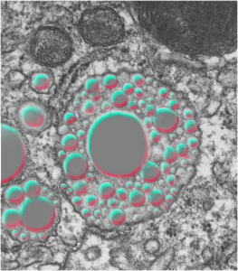
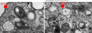
The bottom line of this image is that these droplets appear to migrate along the membrane systems.

perfluorochemicals E2 droplets in murine alveolar macrophage

Some perfluorocarbon based blood substitute – “trivia” or not so trivial

The febrile reaction to PFC based blood substitutes was purported (Donat R Spahn, Blood substitutes Artificial oxygen carriers: perfluorocarbon emulsions. Crit Care. 1999; 3(5): R93–R97) to be related to particle size of the emulsified infusion fluid. Lower particle size was stated to reduce the uptake by the reticuloendothelial system, which increased tolerability, as that stimulation of phagocytosis, and what must be (unmentioned) feedback loops, was deemed responsible for fever and flu like symptoms in patients receiving the blood substitues.

Spahn quotes statistics for IV administration of Oxygent “Intravascular half life is dose-dependent, and for Oxygent was found to be 9.4 ± 2.2 h for a dose of 1.8 g/kg [4]. After the initial uptake of the perfluorocarbon emulsion into the RES the droplets are slowly broken down, and the perfluorocarbon molecules are taken up in the blood again (bound to blood lipids) and transported to the lungs, where the unaltered perfluorocarbon molecules are finally excreted via exhalation. At present no metabolism of fluorocarbon molecules is known in humans.”

I didn’t see any micrographs which gave clue as to “break-down” of droplets, so this is confusing.  I have seen completely solid “footprints” of fluorocarbons, round and with lysosomal enzymes, sometimes a lot, surrounding or off to one side of a droplet, I have also seen the frothy huge lysosomal inclusions which do not have the orderly rounded appearance.

Perhaps just a little overzealous in praise of PFC-based blood substitutes

perfluorocarbon based blood substitutesHere is a quote from a chapter in “Blood Substitutes” (By Robert M. Winslow) and the chapter was written by Jean G. Riess, PhD and Marie Pierre Krafft, PhD. Please note that the location of the primary author is San Diego (home of an ailing Alliance Pharmaceutical Corp Oxygent PFC based blood substitute development) which (like an earlier post on an article by a researcher there – possibly the CEO) also read more like an advertisement extolling the virtues of a product they wanted promote than a scientific pronouncement outlining the effects, both good and bad, of PFC based blood substitutes. I quote one paragraph which is particularly off base, here ‘verbatim’.

Finally, PFCs are not metabolized, and no microorganism is known to feed on them – since Mother Nature did not exploit the PFC route, she did not develop the enzymes that would have been needed to recycle them. Pure PFCs have no effect on cell cultures either, other than the benefits that result from O2 or CO2 delivery. One can drink PFCs by the liter without side effects other than wet pants due to extreme spreadibility!

I take exception to several things here, firstly I have witnessed (with electron microscopy) in vitro responses in the reticuolendothelial cells to PFC:  1) and there are differences in PFC-induced responses, which vary with the PFC, and there are changes in the phagocytic cell population involved, including phagocytosis and the appearance of immune activation. I have seen macrophages that have engulfed enough emulsion particles so that they become half of the visible cytoplasm.  If this is “no effect” I will laugh — it certainly is an effect.  2) also, macrophages  produce visibly increased amounts of lysosomal enzymes that get piled into their phagolysosomes… as very dense caps.  Were that not enough, the last several posts on PFC and macrophages (in vivo) lysosomal inclusions have shown that a huge response in the multivesicular bodies/late endosomes/PFC inclusions to the PFC E2.

Image: cut and pasted and highlighted red cells, shadowed, and also wikipedia’s image of the molecule for perfluorodecalin, the most commonly used ingredient in many of the blood substitutes.  (BTW, none of the various concoctions really were viable substitutes).

MVB/LE/PFC: multivesicular bodies, late endosomes, perfluorochemical inclusions

Alveolar macrophage from a mouse lung, the latter having been subjected to complete submersion in E2 perfluorochemical for three hours, and allowed to recover for 48 hours.

A single macrophage has almost all the PFC inclusions with high concentrations of enzymes, making them appear to have black “caps” in some cases, and in others, mostly enzymes, showing the tiny E2 particles and larger particles (does anyone know if this is Oswald ripening…. it seems as if the particles are doing two things, getting smaller and coalescing…??). The variety of multivesicular bodies here is quite amazing, with some having other structures included, some being filamentous, mostly being very very electron dense.  Each of the MVB/LE with PFC inclusions (i did not tag them all, it was too cluttered) have a bounding box indicated on the unretouched first image,  they are all enlarged and collected in a gallery below (numbered so you can find them).  Compare the variety here with three other posts on MVB/LE/PFC.