Category Archives: Layered intracisternal protein granules in mammalian lung

For 35 years I have thought about this protein organized so nicely in the type II cells of the lung of three mammalian species (guinea pig, ferret, mongrel dog) which I am sure is present in more species, and have not had the time to investigate it further. A peer review publication in the early 1980s was about as far as the study went, though published in an OK journal (Ultrastructure Research) I felt there was more to see than just what I could put down in those few pages. Life makes us make choices, and at this point I choose to play with this old problem. I am not professing anything except diligent “musings” here.

Rabbit alveolar type II cell: is this surfactant protein A or not?

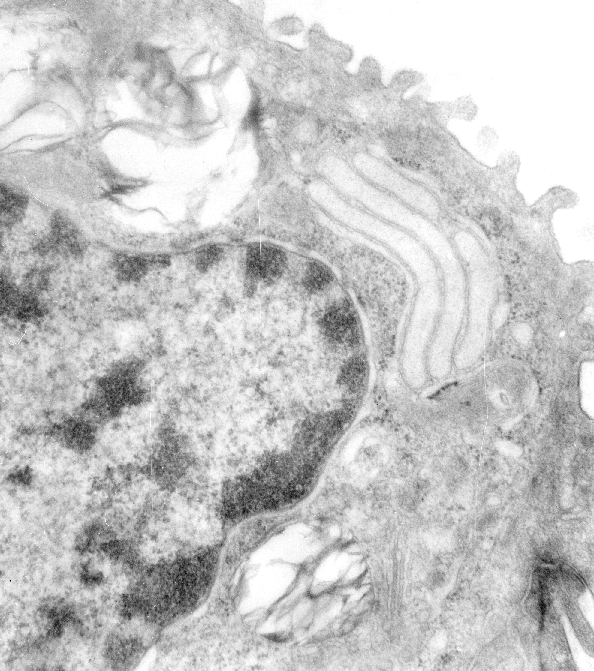
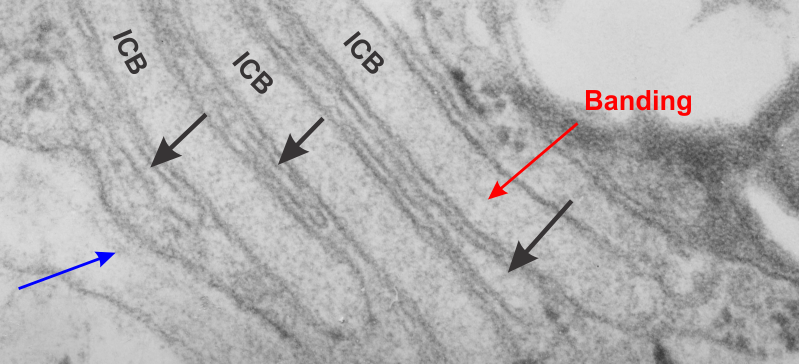
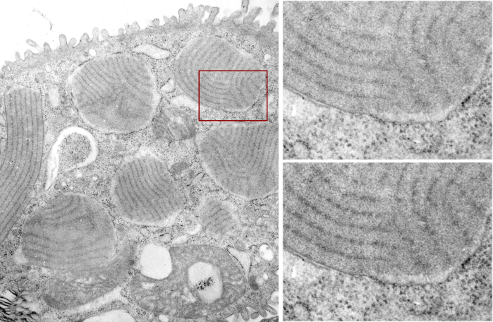
Going through additional species of animals looking for possible corollaries for the intracisternal bodies found in the RER of guinea pig, ferret and dogs, I examined just a few transmission electron micrographs of rabbit. One animal from a study with Leland Clark Jr, very likely involving artificial blood, and four other rabbits in studies with David Warshawsky, with carcinogens (likely dibenzcarbazol, and also iron oxide).  In the former (see the unretouched, but contrast enhanced micrograph below) I did find three stacked RER cisternae which had the look of some of the cisternal bodies found in three species but I was not able to detect any of the layering–aka—periodicity within these cisternae.  Well actually i was tempted to see it, it seemed to me that very faintly there was some tendency to see banding.  You can judge for yourself. Top micrograph.

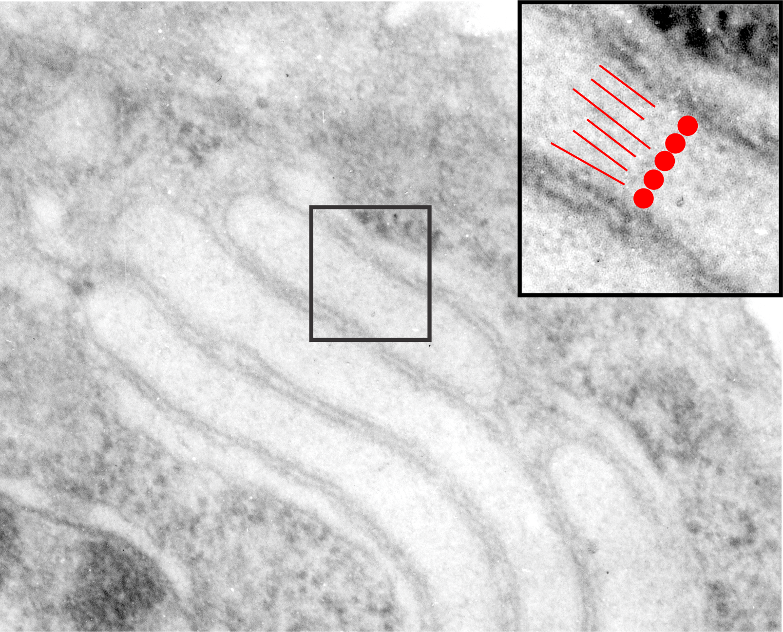
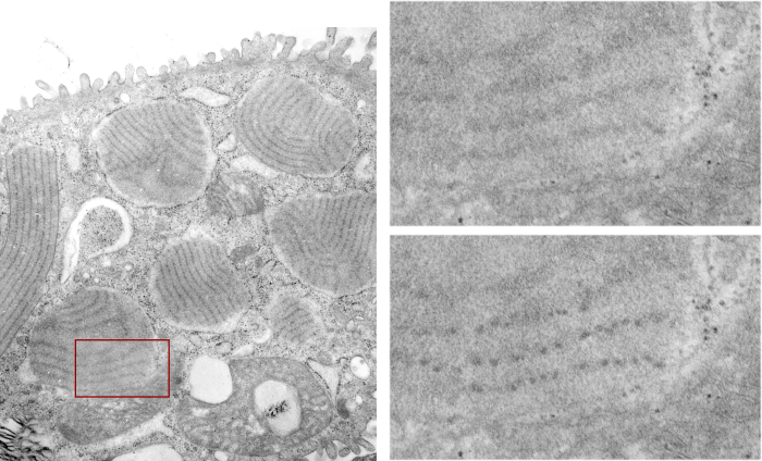
In the bottom micrograph at the thinnest part of the cisterna (inset marked by a black rectangle and enlarged to upper right corner) at a near perpendicular cut, it seemed to me that the cisterna was more than 100 nm in width.  (A ribosome adjacent to the cisterna was used as a measuring stick, the latter presumed to be 25 – 30 nm in diameter;  red dots). What might be construed as linear patterning, or parallel periodicity, appears as red lines.

11320_37150_rabbit11320_37150_rabbit1

Biggest alveolar type II cell RER granule so far

Guinea pig (now the most posted alveolar cell-RER granules (intracisternal bodies) in history (LOL) guinea pig # 301.  This is a very very large protein accumulation, and somewhat different in morphology than the typical intracisternal body (seen adjacent, below) and I don’t know yet why the difference in structure.  This guinea pig, that is, the guinea pig 301 which continually showed the greatest number of RER granules of any other animal in this archival set of micrographs, and interestingly it possesses some distinct prismatic (rhomboidal) regular bodies that resemble virus particles (more data needed).  It would be awesomely consistent with what is known about surfactant protein A and innate immunity to have this guinea pig demonstrate first hand what kind of an increase in surfactant proteins (either A or D) to respond to a viral infection.  We will see.

This granule rivals in size the nucleus in the adjacent alveolar type II cell.

9817_17084_gpig_301_typeIIcell

Osmium tetroxide fixed alveolar type II cell, dog, intracisternal body

Going through archival electron micrographs of alveolar type II cells has been an interesting study. The purpose of the review is to gain insight into what is the explanation for some very interesting, highly organized granules (protein accumulations) that are found in some alveolar type II cells.  Of about 8 species examined so far (rat, mouse, owl monkey, rhesus monkey, dog, ferret, guinea pig, hamster  – even one human case) these bodies have been found in only three species. Dog is one, though in dog they are quite rare, and very small and not nearly as developed as found in ferret and guinea pig.

Most of the archival electron micrographs are tissues fixed in a double aldehyde fluid, in various buffers and at reasonably constant concentrations of glutaraldehyde, around 2.5%.  This tissue however was fixed in osmium tetroxide… thus verifying pretty much that the cisternal body granules are NOT just an artifact of aldehyde fixation.  So this image of a dog alveolar type II cell is not that great a micrograph, but it does show a small, typical style dog intracisternal body granule, with a similar periodicity and banding as found in aldehyde fixed material.

Upper image shows the mostly unretouched (i changed contrast, and took out a few scratches but did not change any data) and the bottom is a highly contrast enhanced crop of the upper image, and i have used the burn tool in photoshop to highlight the periodicity of the middle band (as also seen in many other micrographs posted in this blog).  The ribosome standard I called 27 nm in diameter, which makes the dotted periodicity of the central band of the cisternal body about 17 nm in diameter (also shown as a red dot) and the space between those 17 nm dark protein dots is a distance (made from 12 adjacent measurements) a mean of @ 32 nm.  Band across the body is close to the 100 nm that has been found in other instances in ferret and guinea pig and is presumed to be a collection of some multimer of surfactant protein A.

dog_osmium_fixed_icb


Nuclear portion of an alveolar type II cell enlarged to show nucleoplasm organization

6760_17082_gpig_nuc_pattern

Looking at an electron micrograph of a guinea pig alveolar type Ii cell, (my guinea pig number is 301 – block 17082) archival material from the 1980s, I found this really interesting pattern in the nucleoplasm just above a nuclear pore.  On either side of the outer nuclear membrane spanning the nuclear pore was the granule protein stuff that I have been working on, trying to place it possibly in the surfactant protein family, but being overproduced to the extent that it becomes its 18-mer, in mirror and/or vertically flipped position, times 2… producting the 100 nm banding that I see.  But for this nucleus, just to the right of the nuclear pore marked with a two headed arrow pointing to the lime green intracisternal protein within the RER, there is a pattern that just “popped out” to me which is quite large (relative to the size of a ribosome…. and NOT to be confused with the hexagons in a tangential view of a granule but in the NUCLEUS proper) and sort of hexagonal in shape (which is what one expects when pressing together spherical bodies which I pseudocolored a salmon peach.  Red circles surround structures that look like perichromatin granules (I am sure I could have found more).

Intracisternal granule from alveolar type II cell of the guinea pig

Nucleus, other organelles, nuclear pore, intracisternal protein accumulation, perichromatin granules, and part of the basement membrane are labeled in this electron micrograph of a type II cell from the lung of a guinea pig. Nucleus is on the right and a very thin band of cytoplasm is present, then there is the extracellular space (with basement membranes and parts of other cell processes).  Basement membrane – purple arrow and label; red arrow and label – nuclear pore; black arrow and label – intracisternal protein which might be surfactant protein A (overproduced). Perichromatin granules (adjacent to heterochromatin) are shown by blue arrows and label.

6760_17082_301_gpig

Smooth ER between RER lamellae in guinea pig alveolar type II cells

This electron micrograph shows three very long (a portion pictured here) lamellae of RER with a surfactant ?? protein, maybe surfactant protein A accumulation which shows a very very light area of banding – ICB) , and sandwiched inbetween these three are flattened cisternae (black arrows) which do not apparently contain the protein accumulations that is present above and below each.  Whether these flattened cisternae without apparent protein are going to be sectioned down the block as thickened lamellae with protein content, is up for grabs, but likely (see lowest black arrow which points to a thickened part of the flat cisternae).  This configuration is a little unusual.  Other things to observe in this electron micrograph are: part of a lamellar body – upper right;  tiny hint of the central band typical of what I think is overproduction of surfactant protein A – red arrow; basement membrane beneath the alveolar type II cell, and what is probably an endothelial cell – blue arrow;

icb_10254_gpig

Fuzzy protein granule in rough endoplasmic reticulum of a type II cell

Original, and unretouched or processed (not even contrast enhanced) photo of a small profile of rough endoplasmic reticulum from the alveolar type II cell. It has two ribosomes over to the left about 10 o’clock, they can give you a feeling for the dimensions of the fuzzy balls within the central portion of the profile.  The electron lucent area surrounding the dense center is pretty common in these intracisternal bodies (granules if you prefer) and the layering here is too large to be part of the 100 nm pattern seen when the bodies are found perpendicular to the periodicity.  I looked closely at the center, and I would estimate that the bodies (in groups of three – yes, my guess – roughly) is something around 60-70 nm, at least bigger than the ribosomes (when lumped together in threes or whatever the cluster contains.  OF course the whole granule is much bigger, but it would be fun if the smaller portions of the central density of this intracisternal body equated to 50 – 60 nm groups of three bouquets of surfactant protein A.  That would be just fun.

fuzzy_balls_single_all_grey

You can find a short animation with text at this link to YouTUBE:

More periodicity seen in RER granules in alveolar type II cells

More periodicity seen in RER granules in alveolar type II cells from a ferret. In the areas of this single intracisternal RER protein there are great examples of the periodicity of the protein as it was fixed (this fixation was standard paraformaldehyde glutaraldehyde fluid). This particular electron micrograph is a little grainy (I probably used dektol in stead of microdol to develope it — in hindsight not a good shortcut to take, but sometimes we do things without knowing the 30 year consequences.. duh.  At any rate, I have compared areas in the dense band with the lucent bands of this particular tangential cut through a curved RER granule also called intracisternal body and shown the unretouched (in terms of the densities) and the burned densities as I could see them.  There is a portion where there is not periodicity taken from the exact same RER granule less than 100 nm below the string of beads seen above it.  Ribosome (20-30 nm? is used to compare sizes, and the beads look to be something on the order of 50 nm.

Full granule is pictured in the transmission electron micrograph on top, arrows point to the horizontal segment which has the unretouched periodicity, the arrow below points to the lucent zone used to compare whether I am hallucinating the periodicity (ha ha).  Micrograph 2, just beneath the full granules is enlarged unretouched (i did remove some dust and a scratch using the stamp tool in photoshop but in the line of granules). Micrograph 3, is a view of the exact same strip of the intracisternal body as image 2, but i enhanced the beads with the burn tool. and micrograph 4 is the more or less no-granule-zone in the lucent area as spread out in a tangential cut, unretouched.

more_p[eriodicity_RER_proteins

Ferret alveolar type II cell tangential periodicity in RER granules

More ferret alveolar type II cell tangential periodicity in RER granules. The periodicity is easy to see. The diagram is arranged as previous post, the whole cell on the left, the red box shows the insets on the right, and upper right has the original unprocessed, and the lower right is “burned” in the areas that I think are indicative of spaced molecules (hopefully this is surfactant protein A).  I am open to discussion on this.

icb_6711-23303_more_periodicity

Ferret alveolar type II cell RER inclusions (layered protein granules)

Ferret alveolar type II cell RER inclusions (layered protein granules) appear as evenly stacked bodies, or often on tangential section, areas smeared out concentrically or linearly. These intracisternal bodies, which i think I should begin to call surfactant protein granules (ha ha, not to rile up any feathers) have many subtextures and periodicities which show up when I examine my stash of electron micrographs close up.  Bless my scanner and photoshop, as they allow me to crop and highlight and darken and make what I see initially, into something that can be highlighted for everyone to see.  Such is the case with several of these intracisternal protein surfactant granule bodies found in a ferret alveolar type II cell. The original cell is shown in the video, and a small inset is enlarged, and the periodicity of the protein (which I think is an accumulation of surfactant protein A — maybe upregulated in production as an immune response? I don’t know) along the dense bands of the 100 nm periodicity.  The lighter bands don’t really show up very well on tangential sections, though I have some micrographs where some substructure is visible. The images used to make the video below is here as a png file, where the enhancement of the periodicity can be seen in the figure in the lower right, the area from which this inset is taken is shown in the red box on the left. The entire ferret alveolar type II cell is on the left, apical border facing top.  There are at least 6 inctracisternal body surfactant A granules? present, some showing parallel  substructure other showing curved and tangential cuts.

icb_6711-23303_tangential_periodicity

Here is a short animation using these transmission electron micrographs