Category Archives: Electron microgaphs of lung

Various species of mammal and maybe a non mammal now and then have been obtained and examine using routine transmission electron microscopy. These are summary images.

Before you pitch your old electron microscope –

Before you pitch your old electron microscope -consider these observations.

Firstly, the human brain is a marvelous (and anticipatory) machine. You can scan at break neck speed past grid bars and open space (as one has to do when one is doing electron microscopy on the lung when it has been fixed by inflation) and the section goes by in focus.  That is an amazing computational skill.  If you want to waste time, scan your grid in a newer digital image scope (I used an Hitachi this morning) and i wasted half my scope time waiting for the monitor to update the image so i could tell what i just passed.  Scanning by space and grid bars to find what I wanted to look at closely, and the computer is so slow at updating the images, made me realize that waiting for smear after smear of grey blur took up an inordinate amount of time andleft me feeling like i would have been better off with my old binocular viewport and my old seimens 1A microscope.

Secondly, while the function of automatic bright-dark adjustment is a nice though… again, it is very very annoying. My eyes and brain don’t need to make those contrast changes, they are automatic, and instantaneous. The lighter tissue next to a black grid bar means manual adjustment there, and the second you move more central in the grid space, it changes again.  Not to reiterate the obvious, but the brain does a tremendous job of adjusting, and filtering, and adapting…..  this scope didn’t even come close.

Digital image procurement on transmission electron microscopes (at least this Hitachi at children’s hospital) is a big time gobbler, and is still very very promitive.  and certainly as viewed on the monitor hold no candle to film.

Thirdly, time, how much wasted time. This was supposed to be a time saver, but it recall it took about 14 small steps to capture an image on film with the Elmiskop 1A, but in fact it was less than a few seconds to perform all tasks, on this Hitachi I wait for the final image, i wait for the menu to save comes up, i hunt for the keys in the dark to label (or not label) the image, I wait for the screen to refresh.  It took me less time to write the grid and block numbers in a log book and snap the picture on my antique electron microscopy.

I remember during one “annual review” when an unnamed director was in charge and he mentioned to me that i wasn’t doing enough to get new equipment…. he gave me a bad annual review…. i think because i told him it wasn’t the scope that limited microscopists, it was they 2$ eyes.  I like technology…. but not for the sake of technology.  I like technology when it is an improvement.

I will take the old baby on the left any day of the week.
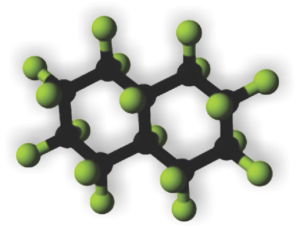
 

PP5 (perfluorodecalin) liquid breathing mouse lung for 1 hour with 1.5 hours recovery

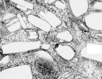
PP5 or perfluorodecalin (image to right) was used as a possible component of artificial blood, or called otherwise, blood substitute(s), but never really attained full research or understanding necessary to determine whether it was a safe substitute for blood or not. Some of the early electron microscopy, which would have benefited from a huge influx of money, early on, for microscopists to examine the effects on all cell types, in vivo and in vitro, but too little was forthcoming to give it a thorough exam. It was clear however, that macrophages and the immune system were going to be big players, both because of the volume of the chemical that was infused or “breathed” and because of its alleged inert nature but odd surface tension properties.

In retrospect, looking at tissues which were taken from Leland Clark Jr’s lab in the 1970s, these particles (droplets) behaved strangely, seemingly dependent upon chemical properties, and of all examined ONLY one perfluorochemical had a shape in the cell other than very very round (this was perfluorodecyliodide – looking like a crystal instead of a droplet, as seen in this image from the liver after infusion of an emulsion).

Liquid in the cell in almost always is “slightly out of round” the best examples being fat droplets. They can be oval or indented or squeezed, as well as round.. but perfluorochemical droplets are ROUND. I would think this could be attributed to their heavier than water density.
Another property (mentioned in previous posts) is that they have various capacities to be re-emulsified, at least that is what i call it, within the cell. Example from the liquid breathing mouse posted yesterday (breathing silicone oil vs breathing E2) shows that the respective macrophages respond by producing different quantities, and also likely types of lysosomal enzymes, in addition to the recycling they do of the alveolar contents (that is, in recycling surfactant and other junk in the alveolar space.

So comparing silicone oil, E2 and PP5… seen here, once again, the lysosomal responses are quite different. Silicone oil didn’t encourage lysosomal production (and surfactant recycling) as much as E2, and E2 in turn caused a lesser (though still pretty dramatic) response than PP5. Actually, from notes taken from this series of experiments (Clark never did anything twice which made it really tough to keep notes) those mice breathing PP5 did not fare well, most did not survive as long as those breathing some other of the perfluorochemicals. E2 was probably the best in this regard, just due to the sheer number of samples and micrographs, this is what I remember.
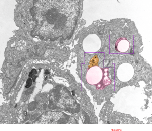
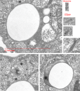
So here is an electron micrograph of an alveolar space from a mouse which breathed PPF for 1 hour and was allowed to recover for 1.5 hours. (1499_5512_PP5_1hr_1.5hr recovery).

Highly rounded droplets, with very well defined lysosomal enzymes. The two non-highlighted droplets are likely to have (as the upper left already indicates) masses of lysosomal enzymes in a different plane of section. There are some smaller, and also very tiny droplets of PP5 visible in the two highlighted LE/PFC bodies. I did find some dense areas of the ER (not likely RER, but something different) that showed an unique substructure (see the inset with three small sample images (enlarged from boxes in the top figure) showing what I noticed.


Inset is enlarged and micron marker is determined from the size of an average (27nm) ribosome, and segments of the inset are all at the same enlargement from the original.

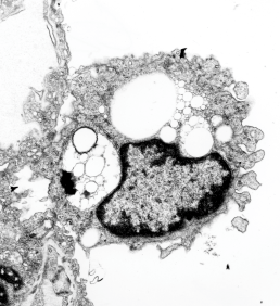
Silicone oil liquid breathing, one macrophage

Here is a not-so-hot electron micrograph of an alveolar macrophage, but the inclusions of silicone oil are kind of interesting. By 72 hours after just one hour of breathing silicone oil, show droplet inclusions that look a lot like they are doing the Oswald ripening thing, but also havn’t stimulated quite the lysosomal enzyme thing that macrophages that have phagocytoses perfluorochemicals show.  There are also bizarre areas of the lysosomes which are not typical of the PFC inclusions.  So differences – noted!

One of the more interesting differences (well two) is that there is almost no discernible trilaminar membrane around the inclusions (LE/SiO droplets) and there is certainly not a double outline (one is the trilaminar membrane of the lysosome, the other the protein coat as seen on PFC droplets (see previous posts)), so those two differences in ultrastructure plus the lack of a definite enzyme overload in those particles in alveolar macrophages that contain silicone oil droplets, and the sloppy separation (or ripening) of the individual droplets of silicone oil vs PFC droplets, makes for nice contrast between the two.

electron micrograph of alveolar macrophage from animal that breathed silicone oil
Here on the left is an enlargement of silicone oil inclusions in an alveolar (1 hr breathing 72 hr recovery) macrophage and on the right, E2 inclusions 3 hr breathing 48 hour recovery (biasing the E2 in the direction of less organization, but still showing much more organization and lysosomal enzyme content than the silicone oil).

comparison of ultrastructure of silicone oil droplets and E2 in alveolar macrophage

Protein coat? in phagocytosed E2 inclusions in alveolar macrophages?

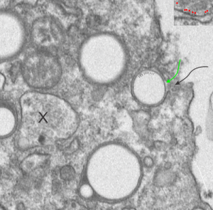
The bounding electron dense coat of proteins on the perfluorochemical droplets, whether phagocytosed from a neat liquid as inhaled from liquid breathing, or provided to the blood stream as artificial blood, it seems to be a fixture of the morphology.  I have scanned the edge of a single alveolar macrophage for evidence for “point of entry” of a droplet of E2 into said cell.  It is pretty clear even from this slightly too thick, thin section of a mouse lung (1349 4840 E2 3hr 48hr recovery) that the electron dense coat around droplets exists in lung of liquid breathing mice before they are phagocytosed by alveolar macrophages.  In this case the proteins likely to be lipid molecules derived from surfactant.

I suppose it is also likely that the slight lipophilicity of perfluorocarbons means that the surfactant lipids (such as phosphatidylcholine and phosphatidylglycerol) and the amphipathic proteins in surfactant (like SP-B) might contribute in some way to this droplet coat (as derived from surfactant already in the alveolus) reflecting its contribution shown in vitro studies suggesting an ability uptake of bacteria from the alveolar space (the leap is to E2 droplets) (ref Yang et al below)

There are at least 4 whole E2 droplets, and two cut off (left hand side of micrograph) droplets of E2 in this image. The tiny dotted periodicity were shown by lines in the droplet at the middle right, the green arrow points to the electron dense bounding layer of “some lipid and protein components from the alveolar space, likely from surfactant”, and the black arrow points to the space in the plasma membrane where the particle is likely being engulfed by the alveolar macrophage. Ribosome size is judged by the upper right corner inset with a portion of RER enlarged identically with the larger micrograph.  X marks an inclusion type that I have seen before but do not recognize. Any suggestions from the audience are welcome (millermn – ucmail dot uc dot edu).

transmission electron micrograph of alveolar macrophage and E2 perfuorocarbon droplets

Surfactant protein B and lipids look like a good candidates for the electron dense cover for E2 droplets — reading the article (Hawgood et al) and Yang et al  specifically referring to SP-B and from which I quote: “In no rank order these activities include  membrane binding, membrane lysis, membrane fusion, promotion of lipid adsorption to air–liquid surfaces, stabilization of monomolecular surface films, and respreading of films from collapse phases.”

SP-B has some interesting properties as referenced in many articles in PubMed which suggest that it has saposin like properties. Wikipedia identifies saposins thusly: “Saposins are small lysosomal proteins that serve as activators of various lysosomal lipid-degrading enzymes”. It is interesting that the huge lysosomal response that is induced by liquid breathing both in alveolar macrophages (and also when perfluorochemical emulsions when given IV) might be seen in the types of lysosomal bodies found, the length of time required to degrade/and/or reassociate the enzymes that appear to cover the droplets in the alveolar space, and concentrate (add more? or recycle more?  possibly be reflected in the fine granularity of the electron micrographic views of these MVB/LE/PFC organelles. The amount of re-emulsivication of phagocytosed E2 in alveolar macrophages appears to be time dependent, and would fit with the tim required to increase in production of some lysosomal enzymes and become associated with the E2 droplets.

Variety: the whole spectrum of lysosomes/inclusions of E2 in an alveolar macrophage

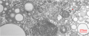
Variety: the whole spectrum of lysosomes/inclusions of E2 in this micrograph of an alveolar macrophage (at least the whole variety I have seen so far). It includes lysosomes (LE/LY/PFC) which probably could be classified all the way from golgi vesicles to late endosomes since it seems that PFCs in general are able to work their way into the whole ER system (though to date I have not seen anything that looks like a PFC droplet in any ER which has ribosomes….  still looking). There are droplets in a “hollow” meaning no lysosomal enzymes yet, on the right but still having E2 droplets with the coating they must pick up from somewhere (maybe alveolar surfactant in the alveolar space?, being lipid and E2 being slightly lipophilic), to the dense small lysosomes with many E2 droplets on the left, to a single E2 droplet on the lower left, and a dozen droplets which look like they are hanging out in the cytosol with no membrane boundaries, to bottom center structure which looks like two E2 droplets in a lysosome with a lamellar type surfactant inclusion.

There is no paucity of free ribosomes in this cropped image, and size can be inferred from the approximate 27nm diameter of a ribosome (red dot), to the larger droplet size (bar=270nm).

Countless PFC droplets in alveolar macrophage lysosomal body

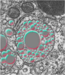
The lysosomes in this alveolar macrophage from a mouse which breathed E2 for 3 hours and then allowed to recover for 5 days are awesome.  The macrophage has produced enzymes which re-emulsify the E2 into very small (in most cases) droplets within the lysosomal structure. In addition, the enzymes make a border, which is very difficult to distinguish from the trilaminar membrane which surrounds the lysosome proper.  I don’t know how to explain this look of a double membrane, but figure it is partly a physical interaction between the E2 droplet (and not unique to E2 but seen with many other perfluorochemicals) and its hydrophobicity, and slight lipophilicity.

The smallest spheres of E2 are down around 35nm diameter, and size can be compared to something just larger than a ribosome (@27nm). Picture on the top is the unretouched (i may have removed a scratch with the band-aid tool in photoshop but nothing else), and it is not too great an image (scanned from the 31/4×4 acetate negative), but interesting still, and providing lots of information. Image below is one where I have highlighted the E2 droplets in a single membrane bound lysosome and embossed them.

The dark structure (also rounded) in the upper right hand corner of the images is what I think is a phagolysosome that contains mostly phagocytosed surfactant lipids (some layering and myelin-look is seen within this structure) from the alveolar space. There are also two mitochondria near center top. It also seems likely that some of the surfactant engulfed by alveolar macrophages would find its way (during re-purposing or re-cycling) into such lysosomes containing E2 droplets.

electron micrograph alveolar macrophage E2 liquid breathing perfluorochemicalelectron micrograph alveolar macrophage E2 liquid breathing perfluorochemical PFC highlighted

Is this E2 in alveolar endothelium?

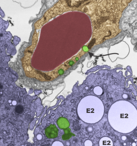
Loooooking over an old negative of lung from a mouse that breathed oxygen bubbled liquid perfluorochemical  (in this case E2, which is a freon) I spotted what could be, maybe, seems almost possible, droplets of E2 in the endothelium (alveolar sac endothelium).  I selected portions of the image out and pseudocolored them for easy identification: Red cell within the capillary lumen, red, endothelium orange, lysosome/or vacuoles that might be E2 and comparison with those which are in a nearby alveolar macrophage, green, and the macrophage cytoplasm is purple. There are some reasonably large E2 droplets which are probably unequivocal (is that not an odd combination of words?) with more distinct membrane boundaries, and absolutely NO fuzziness within the inclusions but also having the distinctive “black cap” of lysosomal enzymes off to a little blip on some side or other of the droplet.   This is NOT seen in the endothelial inclusions…. I will look for less equivocal samples but the similarities between those and additional ones found in the macrophage is clear. 1351_4840_mouse_E2 breathing for 3 hours, 48hr recovery, capillary_endothelium and two macrophages.


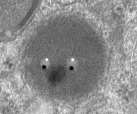
E2 lysosome “Looking at U”

Could not help but post this cute little lysosomal structure in a macrophage of a mouse that liquid breathed E2 (see previous posts for chemical structure) for 3 hours and was allowed to recover for 48 hours. This lysosome is a tiny portion of an electron micrograph of a macrophage in the alveolar space of the lung.

The scope of lysosomal variation in these macrophages is really quite astounding.  I see lysosomes with a tiny bit of enzyme material localized on one side of the membrane bound structure, and others, like this, with maybe 4 or so tiny perfluorocarbon droplets (which looked like two eyes to me), and massive amounts of enzyme (all that medium grey in a round membrane structure). A library of these variable organelles is what I am attempting to do at this point.  E2 is capturing my attention. If you look closely, around the periphery of the lysosome there are small circular areas, which I have no doubt are the tiniest lysosomal droplets down around the size of 20-30nm in diameter.  The “eyes” that are droplets here are much larger than that, closer to 100-200nm). Highlight in the eyes and the dark pupil i did in photoshop just to have fun, but the eyeball itself is E2 and the “nose” is enzyme density.  In the upper left corner of the image there is another lysosome with a round E2 droplet (about 75nm diameter) with some layered darker substructure.

It is possible that part of the lysosomal material here is unique to lung since alveolar macrophages (which this is) tend to uptake surfactant in large quantities, and it seems that some of this lipid material (perfluorocarbons have different lipid solubilities, and this might contribute to the mixture of electron dense (osmiophilic) nature of the lysosomes. I might call these MVE/LE/PFC, but that is too cumbersome, hence forth, just lysosome. Besides the specific proteins and enzymes and pH in these structures has not been measured in this archival material… thus there is no way to accurately classify them so I will call them lysosomes.\


Perfluorocarbon compounds and lysosomes: slippery mix

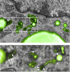
I posted a few days ago (here) a phrase which i thought was a little off color, and definitely made for effect, but stirred up something of interest in the way that perfluorochemicals “slippery and inert” might create or produce unusual lysosomal structures.  I did post a composit lysosomal structure which looked to be like a little pouch full of varying size beads, or droplets, which are presumed to be the footprints of perfluorochemical left after liquid breathing or invusion of PFC emulsions.  I examined some additional lysosomes this morning and it is clear there are many tiny droplets that are mixed in among some very large droplets, and these are so close to the nucleus, and near, perhaps almost looking like a connection, some of the ER in the cell which could be classified as RER.  PFC ispossibly moving back up the ER profiles, and while all the names for endosomes (early, late, hybrid, budding, bla bla) this system is obviously continuous and naming “early” “late” etc  adds little to the understanding (in my humble opinion), but naming them by pH and activities and/or membrane proteins associated, would be beneficial. Another issue is why there is a distinct dark band around the PFC droplets, it gives the appearance of TWO trilaminar membranes rather than a typical membrane bound endosome or lysosome.

Below is a micrograph (actually a scan of the original negative since the print I had marked and draw all over) which has two magnifications. Top image has white box which is enlarged below it, and the long stringy lysosomal structures are very close to the nucleus, even adjacent RER.  this is almost like the “egg in snake” appearance.  Presumptive perfluorochemical droplets (in this case E2 — structure given HERE) are colored green, the most electron dense areas surrounding the green (darker green) are lysosomal enzymes (also presumptive since I have not done any specific staining to verity it… but it is obvious.) White arrows point to the tiniest droplets (some in the range of about 30 nm, and some of those light areas may be tangential cuts off the ends of larger particles.

I suppose it is possible that the movement of PFC up the RER is to be expected because of its spreading chemical properties (at least according to Jean G. Riess and Marie Pierre Krafft.

Not typical lysosomes: Do engulfed PFC droplets migrate up the ER

Looking at lysosomes in the lung of mice which have breathed perfluorochemical (PFC) liquids is interesting. These are old archived micrographs from the early 1970s but have a lot of valuable information about the reactivity of perfluorochemicals in the body (the latter being touted as totally inert.  Regardless of what science says about the reactivity of PFC they do cause a mountain of effects in humans, not the least of which is an immediate uptake by the reticuloendothelial system, massive sequestering and dumping of enzymes into the resultant lysosomes, and a kind of partitioning, or re-partitioning, or re-emulsification of the PFC, depending upon whether it was breated as a neat liquid, or infused as an emulsion with other agents accompanying it.

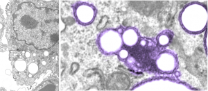
So “case in point” is this alveolar macrophage, picture taken from the lung of a mouse which breathed E2 for 3 hours and was allowed to recover for 48 hours then lungs taken for electron microscopy (and probably chemical analysis, though I have no clue where those data are at this point 40 years down the pike).  I just know that my negative number is 1351, and that block number is 4840, magnification was 5,000 (Zeiss 10) date 2 17 1975 at 60kv.  Mouse weighed 16.85g, breathed E2 for 3 hours on 2 3 1975, and was processed on 2 5 1975, which equates to 48 hour recovery. The sample of lung was taken from the R, mid lobe.

The image below (L) is a portion of the original negative (scanned as a transparency from the film) with an enlargement (box demarcates it) on the right where I have “pseudocolored” the protein densities with purple, which surround the empty footprints where the PFC was fixed (leaving the nice round spaces) in the cytoplasm. About 6 ribosomes (@27nm diameter) are also highlighted beneath the central phagolysosome?lysosome?membrane structure to show that the size of the smallest perfluorochemical droplets is in the nanometer range, before — before what, perhaps being small enough to diffuse back into the alveolar air space and be exhaled.  I don’t believe anyone knows whether the larger or the smaller E2 droplets are more apt to move into the alveolar air space.  There is one tiny droplet which is about 27 nm nesteled between two ribosomes… go figure.  The enzyme response here is pretty amazing, and appears in section to be most often seen as spheres, but is more likely to be a longer sausage shaped lumpy body.

The bottom line of this image is that these droplets appear to migrate along the membrane systems.

perfluorochemicals E2 droplets in murine alveolar macrophage