Category Archives: Electron microgaphs of lung

Various species of mammal and maybe a non mammal now and then have been obtained and examine using routine transmission electron microscopy. These are summary images.

Hamster alveolar type II cell: electron micrograph

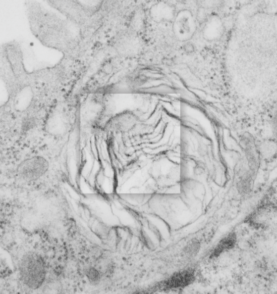
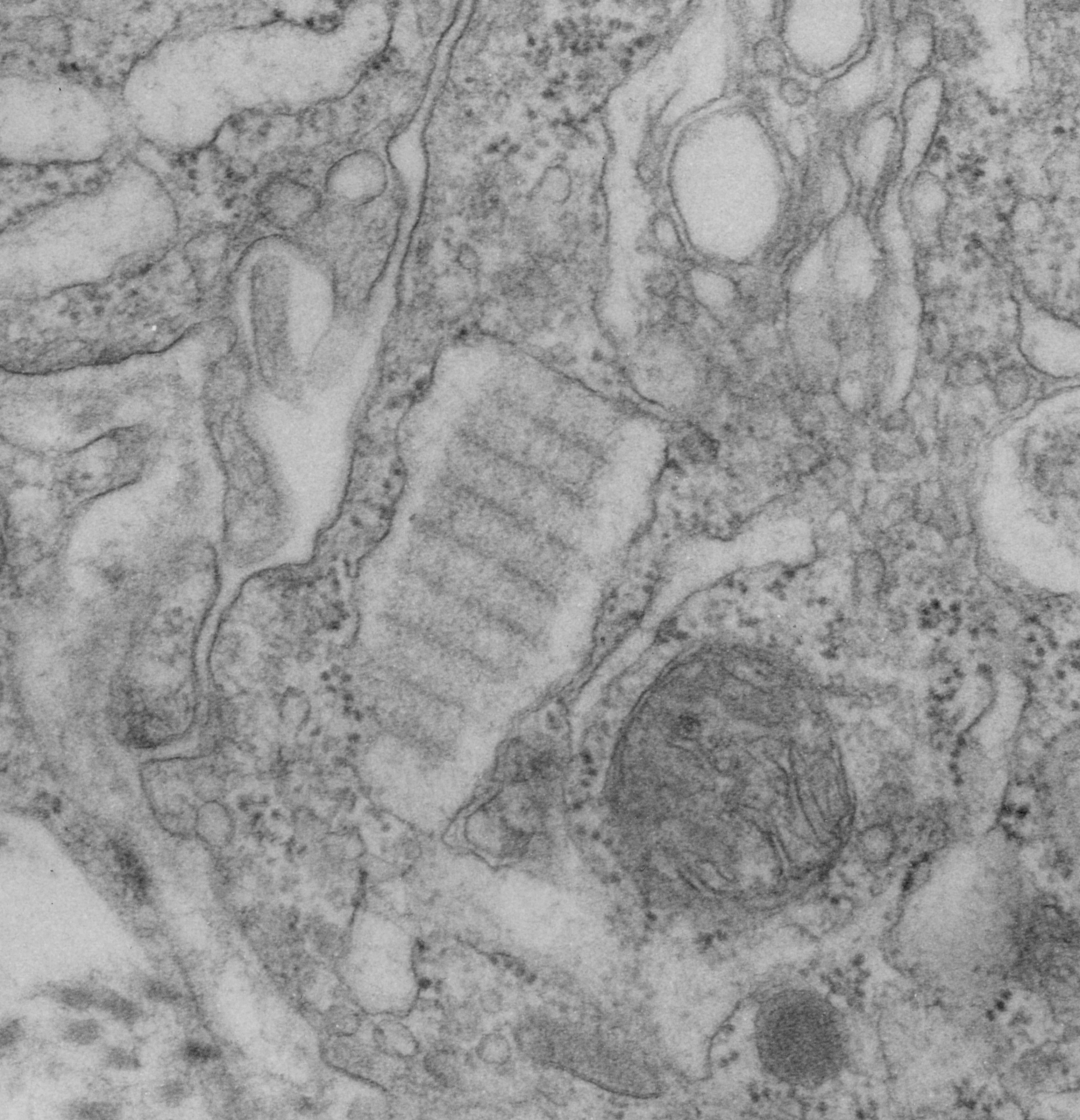
Still searching, here, for clues as to what types of organizational patterns can appear in the rough endoplasmic reticulum of alveolar type II cells. It becomes apparent that not all species have these organizational things, the intracisternal bodies, or granules. The hamster comes close, perhaps, and if this is not my imagination, I have highlighted a periodicity (mostly dotted) within a few profiles of RER in the cytoplasm of an alveolar type II pneumocyte.  There are some obvious differences between this organization (if it is not artifact) and the granules that appear in the guinea pig, ferret, and dog type II cells that are noteworthy. 1) if the dotted appearance is real, then it is perpendicular to the long axis of this RER profile, and 2) if this is a surfactant protein organization at all, then it doesn’t demonstrate the same periodicity as the other three species.  This doesn’t mean of course that it is not a surfactant protein, ’cause it likely is. Even at the widest part between rows of “dots” there is probably not 100 nm…  maybe 80 or 90, so that puts the size a little off.  Ribosomes in blue can be used as a reference to approx 20-30 nm diameter.

7287_24716_hamster_alveolar_type_II_cell

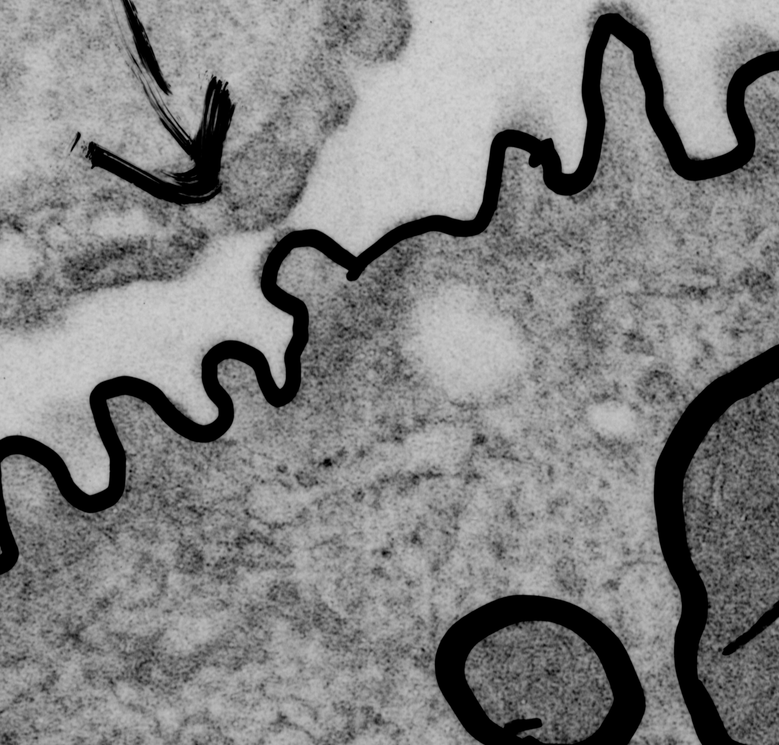
Yellow orange body is the RER profile with dots,  purple-blue, ribosomes for comparison in size.  (neg 7287 block 24716 hamster, born 9 1981, presumably female, animal 2 of three, @ 1.5 yr old.

Lamellar body in an alveolar type II cell has surfactant-like-tubular-myelin grid pattern

Lamellar body in an alveolar type II cell has surfactant-like-tubular-myelin grid pattern. This was found in an electron micrograph of a ferret type II cell (neg 9872, block 23462, ferret # 7, control, 6 10 1982). This particular lamellar body is unusual in that within the lamellar structure, there is a grid like pattern which is normally seen only in the alveolar space. While, according to the literature, alveolar type II cells can internalize mature surfactant from the alveolar space, this doesnt anatomically seem very likely to me as an explanation for this particular structure.  I thought it was unusual enough to post. I put a bounding box and a drop shadow around the area of interest.  The grid inside has at least 4 partitions horizontally as well as vertically.  Surfactant protein A is likely responsible for the grid (according to consensus) though this is usually in the alveolar space. This mineature tubular myelin was described in a book, ed Jacques Bourbon I think, which was reported to be about 1/3 the size of tubular myelin in the alveolar space.

9872_23462_ferret_lamellar_body_grid

Here are two reference images for the above micrograph to give approximate sizes. Regular tubular myelin has a side to side distance of something around mini_tutular_myelin_in_lamellar_body1100 nm, whereas this intra-lamellar-body mineature tubular myelin sample is about 60 nm?  Ribosomes are 25-30 nm (black circles)


Electron micrograph of mouse lung after liquid breathing E2 for 3 hours

Feb 19 1975 under the auspices of the Children’s Hospital Mental Retardation Research Foundation (no longer in existence) house in what was called the IDR building (Institute for Developmental Research – also no longer in existence) with Leland C. Clark, Jr, I was in charge of looking at histological changes from his experimental procedures using perfluorocarbons: as 1) possible components of artificial (or synthetic) blood, 2) for contrast agents in lung, 3) and for lung lavage and aeration fluids.  From the collection of animals and micrographs, here is a portion of a red blood cell, taken from an awake swiss albino mouse who had breathed E2 for 3 hours, and allowed to recover (apparently uneventful) for 1/2 hour.

I am totally not sure if anyone in the world will care about these data, but could not in good conscience allow some of the images to go into the recycling bin without recording them for posterity.  Given long life, I will at some point assemble them into a manuscript, to submit “where”  ha ha. Just relating this to the presumptive SP-A cisternal bodies (granules) that I am working on, none seen in this particular mouse (11 micrographs with type II cells at sufficient magnification to assess the RER and the perinuclear membranes.

Neg 1401, Block 4960, mouse 18.63 g, mag at scope=8,000 x, enlarg=2.83x, Karnovsky’s fixative instilled in trachea, osmium post fix, EPON 812 embedment.  Original description of section with LM = general increase in cytoplasmic lucency, no plately plugs, blood vessels (per this photograph) were mostly open.  Not seen in this micrograph but present in others to come, surfactant hypophase was abundant, the cytoplasm to nucleus ratio seemed increased, no E2 inclusions were found in alveolar macrophages.  Red cell within blood vessel at bottom, endothelial nuclei diagonally across middle of micrograph. Plenty of “transforming fibrils” as Dr. Lowe and Dr. Charles Basom used to call them are seen in the interstitium.  No alveolar type II cells in this image.

1401_4960_mouse_lumg_ E2_pfc_breath_3hr


Alveolar type II cell morphometry: old school

This archived electron micrograph is a sample of the old school style of morphometry. Here is a ferret alveolar type II cell which, when printed, was overlain with a scored plexiglass grid during photographic exposure, to make it easy to count intersection-crossings to determine the volume density of organelles, such as mitochondria, lamellar bodies, RER profiles, and also to count intersections of linea profiles for organelles like RER and plasma membrane and nuclear membrane.  You can see in this very old sample that I circled the nuclear pores.  Cross marks at intersections counted are still visible (ink lines).  Though this is seemingly primitive, to me it allowed much more precision that using image processing, since the eye can adjust to differences in light and darkness in the prints, and pick up debris and other extraneous junk and scratches that might just be counted as “objects” in computer vision programs.  Besides being more flexible, probably more accurate (my opinion of course) it is also quick, and does provide a “hard copy” to go back and verify and spot check one’s data (which counting on a computer doesn’t always do. Even more importantly, not all information that is desired is known at the time of sampling, and therefore having a resource to check back with is invaluable.

4532_18332_ferret_typeII_morphom

Fuzzy protein granule in rough endoplasmic reticulum of a type II cell

Original, and unretouched or processed (not even contrast enhanced) photo of a small profile of rough endoplasmic reticulum from the alveolar type II cell. It has two ribosomes over to the left about 10 o’clock, they can give you a feeling for the dimensions of the fuzzy balls within the central portion of the profile.  The electron lucent area surrounding the dense center is pretty common in these intracisternal bodies (granules if you prefer) and the layering here is too large to be part of the 100 nm pattern seen when the bodies are found perpendicular to the periodicity.  I looked closely at the center, and I would estimate that the bodies (in groups of three – yes, my guess – roughly) is something around 60-70 nm, at least bigger than the ribosomes (when lumped together in threes or whatever the cluster contains.  OF course the whole granule is much bigger, but it would be fun if the smaller portions of the central density of this intracisternal body equated to 50 – 60 nm groups of three bouquets of surfactant protein A.  That would be just fun.

fuzzy_balls_single_all_grey

You can find a short animation with text at this link to YouTUBE:

Odd protein accumulation within type II cell of an aged guinea pig

Ultrastructure shown here of more unusual protein structures within the RER of type II alveolar cells, these are really not at all like what I have been thinking are SP-A, but are more like fuzzy balls.  The dimensions of the rounded electron dense  accumulations within the cisternae of RER within this type II cell are about the dimension of 100+ nm.  The comparison in size between three ribosomes from this same image (see the inset in lower right at actual size) and three of the fuzzy balls? perhaps, in the same inset at actual size, and then see the two types of structures, i.e. the ribosomes and the fuzzy balls enlarged to the same degree and pseudocolored in the inset to compare their relative sizes.  The ribosomes are supposed to be something around 20-25-30 nm in that range, and if these are fuzzy balls (that would be multimers of surfactant protein D) then the fuzzy balls should be about three diameters compared to the ribosomes.  Doesn’t quite work out, so the identification of these electron dense objects within the cisternae of RER in this electron micrograph is up for grabs.  Nucleus is pseudocolored blue, nuclear pore, green, RER profiles with the odd protein, orange, actual size ribosomes and fuzzy balls grey, in inset, and enlarged in relative size, ribosomes, blue, fuzzy balls red.

6776_17082_gp_typeII_cell_ICBs

Nuclear pore: canine type II alveolar cell

nuc-pore__dog_type_II_cell_nucleusThis cropped portion of an electron micrograph of a nuclear pore was taken from the nucleus of a canine type Ii alveolar cell. It shows the filaments on the cytoplasmic side, and a bunch of ribosomes positioned just outside the nuclear pore, and two very distinct structures, about ribosome size that are either coming or going through the nuclear pore.  It made me laugh, as it almost seemed as if they were going or coming in a “pin ball” fashion, being shot through the pore directly in the center. Do you see both of them? one is almost dead center of the micrograph, abd just elow the nuclear pore, and one is almost directly below that.

This electron microscopic image prompted me to try to figure out what is known about the nuclear pore complex in general, and every diagram I found in the literature was just a little different, even that found on wikipedia had some elements that I could not assemble over top the micrograph (which is basically what i wanted to do), so the diagram which accompanies this fine structural view (ultrastructure) is way off… I would love to find the time to put together a better diagram.

nuc-pore_diag


Rough endoplasmic reticulum, ribosomes, and protein synthesis

Pseudocolored electron micrograph depicting what I think is 4 separate actual protein transcription events within an RER profile — see as four ribosomes on the RER membrane and the resulting 4 strands of protein within the lumen of the RER. The RER cisternal profile is pseudocolored cyan, the ribosomes (4 of them in particular) are blue, the proteins being synthesized, 4 of them, from adjacent ribosomes are in red, and the extracellular space beside the type II alveolar cell (from a ferret) is brownish, and the type II cell cytoplasm itself is a light violet color. This was an accidental find while perusing hundreds and hundreds of type II cell electron micrographs from a half dozen species of mammal.

This particular micrograph was not taken FOR this purpose, but it illustrates a point in time frozen by fixative and plastic three decades ago.

9777_34065_RER_ribos_prot_color

Ferret type II alveolar cell RER showing parallel bands in organization of intra-cisternal protein

How awesome is this particular view of an intracisternal protein in a ferret type II alveolar cell. The bands are slightly tangential and so only the most electron dense banding is seen, however, the interesting thing here is that on the periphery there is not really any banding, but it looks as if the banding occurs centrally in the granule (aka within the profile of the RER).  Also, in the upper left of this electron micrograph (in an adjacent type II cell) examine closely the profile of RER  which actually can be perceived as ribosomes on the membrane surface and the trails of protein hanging off in the RER lumen.  Really a classic text-book presentation in “real life” so to speak, though we all know that TEM captures only a “nanosecond in time”, and all proteins are “fixed” and so distorted. Nevertheless, this may be one of those opportune views.

The protein (which I think is SP-A) is central in the micrograph, the banding pattern is at about a 35 degree angle.  Ribosomes are present on right and left borders for the most part, smooth ER is in the upper right corner and there is a tiny portion of a lamellar body off on the right side.  A portion of intercellular space with a couple of plasmalemmal folds crosses from top center to bottom left. There are a few other profiles of RER without SP-A? banding as well.

9777_31065_ferret_crop

Cat: periperhal lung type II cell

Lung tissue harvested from a sham treated control from another investigators experiment (thereby saving time – money – animals) here is a piece of a type II cell viewed with the electron microscope. It has my markings (for morphometry, back in 1982) and a marker arrow pointing to an area of RER which was the closest thing i could find to an organized protein in the RER (as a continuation of the search for species which may have SP-A organization similar to that found in guinea pig and dog and ferret.  In cat, though the total number of electron micrographs I have saved is not that great (a dozen or so) therefore the absence of such a layered intracisternal protein is not to be assumed).  The ribosomes studding the RER membrane provide an estimate of size (each being approximately 25 nm in diameter – a general number for all species and conditions…. (generalities are not always good for much, but an approximate size is OK here).

7296_24554_cat_3_lung