Category Archives: Nuclear pores and nuclear architecture

Nuclear pores and nuclear architecture transmission electron microscopy

Asymmetrical apoptosis with four blips

I ask forbearance for my posting this. I have seen this asymmetrical type of apoptosis in some experiments that were done while investigating an anti-apoptotic gene. I was just doing the electron microscopy, others were doing the cell culture and knockdown experiments.  Typically apoptosis in culture presents with a round nucleus lots of large nucleoli and a bilateral (mostly) distribution of granular material along the inner nuclear membrane of the cell as it dismantles itself. The squeezing and pinching in this type of apoptotic cell was just really unusual and as is always the case on government grants, the reasons were never flushed out because of time and money constraints.unusual apoptosis in knockdown tissue culture experiment A549 cells I bet there is a really interesting reason for these, and this particular set of culture dishes got a scrambled version of an si67 probe and was supposed to be a control. It seems to me that part of the nucleus a just got extruded? I did count 8 nuclear pores (main criterion being an obvious set of cytoplasmic filaments off the cytoplasmic side of the pore.  Few mitochondria, many vesicles. One interesting thing about this apoptotic style is that the cytoplasm is not full of ribosomes as occurs in apoptosis in many cell types.
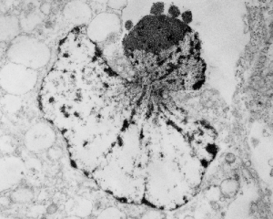
It is not something i could have made up (LOL) but my initial guess is that the four little ball-antennae are some remains of cajal bodies, and heretically i could suggest that the linear divisions radiating out into the “skirt” of whats left of the nucleus, likely has something important to do with each chromosomal territory (separations, walls, scaffolding, call it what you like).
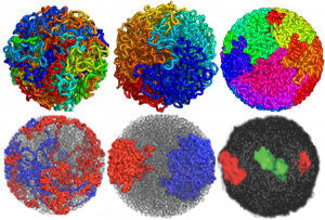
 

Chromosome territories and models of folded DNA

DNA folding: fractal globule and equilibrium globule renditions.  What awesome research and a daunting amount of data to handle. I read (or tried to read) one of the original papers by Job Decker  and another by Erez Liberman-Aiden but could hardly wade through them.  I am linking a blog called Explorable which was easier to read and recaps some of the figures in the original publications.

Altogether awesome, but I struggle that some the images which could actually use real electron microscopic (TEM anyway) structures so show these types of data…they don’t do so yet and there is much that could be revealed.  That would be a good way of bridging what is seen with the theoretical. The radial symmetry (on 2D bilateral using TEM) was obvious to me decades ago, not in the interphase cell so much as in the cell undergoing apoptosis… I am so glad this will be the case).

Here are a couple of images which show different models (fractal and globular) for DNA folding, and one that shows the radial symmetry found in the more_transcribed (which i would dare to call euchromatin, and lesser_transcribed (which again, i would dare to call condensed chromatin) regions of the DNA. I had always kind of thought that the active regions would be at the interface between those two TEM visualized domains…particularly at the nucleolus.

What haunts me still is what I saw 50 years ago in tissue culture is the spinning nucleus (time lapse photography), I have yet to have anyone put that into the puzzle, for a motion and distribution to the nuclear pores, of newly transcribed RNA.  Certainly that is in the mix somewhere, unlike these static models.  Below is a shameless cut and paste of the several images that come up on google for DNA chromatin territories (some fractal globule, some equilibrium globule, far right lower, green is more highly transcribed region of a chromosome.)… AND where is the nucleolus.


Great humor: great truth

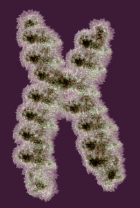
Reading about nuclear organization i ran onto this quote, and felt it was so great i just had to repost it. I personally continue to marvel at the naming of cellular structures, processes, and stuff, and the term “junk DNA” always rubbed me the wrong way, as does “non-coding DNA”.  I marveled to myself when reading those terms “what gives individuals the ego to believe they know enough to call anything “junk DNA”. I am showing a diagram of a chromosome (vector file with thousands of loops, and highlighted edges, iridescent graphic from a failed cover submission trial, a decade or more ago).

Well, here is someone who actually got that into a reputable journal… good for him — quote—from Thoru Pederson. Half a Century of “The Nuclear Matrix”, Molecular Biology of the Cell Vol. 11, 799 – 805, March 2000: “These kinds of ideas have been generally ignored because the noncoding DNA is so “uninteresting” as sequence (as if we were at present clever enough to be able to detect all “interesting” DNA text, which we certainly are not). At our present state of knowledge (ignorance) we can only view the noncoding DNA’s information content on the basis of what is absent [e.g., promoters, cap sites, splice sites, terminators, and poly(A) sites].”

Estimates of the size of some nucleolar components

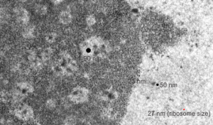
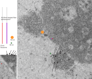
This micrograph, parts of which have been seen before, is an hepatocyte nucleus which is in the early stages of demise (untreated CoS14 ko without rescue). The nucleolus is large, and has two cajal type bodies (one seen here) and fibrillar centers with fairly large densities within (of approximately 90 nm diameter). I have given these sizes (relative to a ribosome size red dot approximately 27 nm (lower right hand side of micrograph labeled as such). The granules within the granular portion of the nucleolus (as well as the banding patterns found on cajal bodies) is something around 23 nm.  What I presume to be perichromatin granules look to be about 50 nm. The large dense bodies within the fibrilar centers are a little unusual.

Interchromatin granule cluster ?

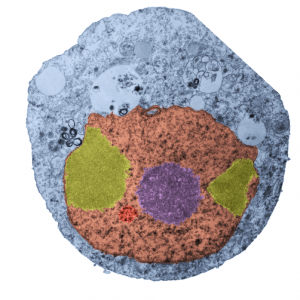
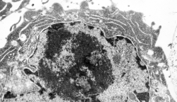
Nothing is black and white, except electron micrographs, and even then they are easy game for dodging, blending, blurring, mending, and pseudocoloring. Cytoplasm here is blue. In this case I have a cell from a culture of A549 cells in which C9orf82 was knocked down using si67.  This is pellet 4 from one of those studies. It produced this cell in which the granular component of the nucleolus was separated into at least two bodies (almost bilaterally symmetrically arranged) which rest at the inner nuclear membrane. Fibrillar centers are small, dense fibrillar component is still visible (albeit not distinctly) in the nucleolus (pseudocolored purple in the center of the cell).  Off to the side is what I could guess would be either degraded DNA (not my first choice as an answer (it is pseudocolored bile-yellow) or perhaps a fine granular component of the nucleolus in an apoptotic cell.  Part of an interchromatin granule (red, lower left) cluster (IGC) (sometimes called speckles, a name which is not really to my liking since the IGC has a boundary, a background texture that is also part of the structure).  So I would propose keeping two names, the “IGC” which is the greater boundary of that area and “nuclear speckles” as well, since the densities (as seen in this micrograph) can apparently be large.

If i am not off base here, the background of the whole IGC is red, but there are very clearly larger than typical speckles within the IGC, and the latter itself is quite small.  With immunohistochemistry the diffuse staining of IGC background proteins fluoresces with some of the antibodies to proteins like SC-37 in an area which can be 2 microns across easily.  I am looking for alternative proteins stained in very punctate regions of the IGC and hopefully they will be different and good markers for a separate ultrastructural components of the IGC. The point would be to localize to the densities with the IGC, some proteins for splicing, and accept that the diffuse staining could apply to SC-37, but not other proteins, mainly those in the variable size granules within the IGC.

We will see if there are data to back up the keeping of both names but assigning them to their obvious separate entities. BTW, there is pretty obvious bilateral symmetry to this apoptotic cell nucleus. and a radial symmetry to the dense bodies within the red colored IGC. And while i have no clue what the curley-Qs are… i bet they have something to do with the transfection. Just looking at this cell shows one tiny surviving mitochondrion, in this apoptotic cell, which is very close to the cytoplasmic filaments of a nuclear pore…also something which needs to be monitored during apoptosis.

Awesome gate: the nuclear pore

In the past 30 years the nuclear pore has been studied, as it is the gatekeeper between nucleus and cytoplasm. It seems to me that it is probably not the only way into the nucleus, and i freely say this from ignorance of nuclear pore biology, but just because in general, it is very unlike “mother nature” to provide only one way to do things.

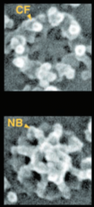
Their figure Figure 2 from which I have cut and pasted a portion, is just beautiful, looking into the nuclear pore (using SEM) from the cytoplasm of the cell, seeing the nuclear pore basket below, and then looking up from the nucleus of the cell outward, seeing the basket in the center.  What I was struck with is the relative emptiness of the areas between the 8 spokes of the nuclear pore, and immediately thought that the transport of small molecules could just go through there..but I would have to think that the pore would be unlikely to let past 10 nm molecules at the same time as a 39 nm molecule (i think that was documented in some study…  especially since the total central pore unit is 45-50 nm.  I can visualize this wacky wiggling of things being imported and exported through the center of the pore bouncing from side to side, letting smaller molecules go through when possible as larger molecules get transported through.  Reminiscent of the activity in a pinball machine bouncing around.


Nuclear pores: diagrams vs micrographs

It is sometimes difficult to reconcile all the graphics that appear in research articles with what actually one sees with the electron microscope.  There are artists and scientists trying to communicate facts and abstract ideas and the results are something hysterical.

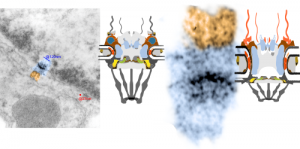
Anyway, in looking at the 14CoS ko 48 hr no rescue nucleus in an hepatocyte I found this nuclear pore which actually had what looks like some of the 8-mer basket proteins that were quite clear.  I have highlighted those (on the nuclear side of the pore) in blue and highlighted the cytoplasmic side with cytoplasmic filaments in rust color.  One can see from a diagram from wikipedia (thank you wikipedia) which I have extensively simplified and modified, that the original and what I have seen in actuality don’t really fit.  I cut the diagram apart and extended the inner nuclear membrane space, shrunk up the basket and extended the cytoplasmic filaments (see diagram on the right).  The other issue is likely that the micrograph tends to show the pore from the ” cytoplasmic viewpoint” or bottoms up, while the diagram looks more “at the horizontal level”.  Basically, the nuclear pore complex is an absolute wizard structure… fun to learn about. This electron micrograph is from a 48 hr old 14CoS ko pup, not rescued with NTBC. 18750_80206_anim#35 liver.  More posts on this topic HERE.

48 hr old 14CoS ko pup, not rescued with NTBC. 18750_80206_anim#35 liverWhat i have trouble visualizing with electron microscopy are the three rings…. the cytoplasmic ring, the nuclear side ring and that ring structure that individuals claim is at the rounded fusion of the trilaminar inner and outer nuclear membranes.

Here is another nuclear pore from the same electron micrograph in which I have highlighted the four areas which perhaps will correspond to the nuclear side ring 2 dots at the bottom (along with light blue arrow of some particle just on the way out of the nucleus likely) and the cytoplasmic side ring above). Orange arrow points to where the trans membrane ring should be seen.

nuclear pore complex electron microscopy

Nucleolus and SV40 infection

Looking at the sum total of nuclear architecture seen from the viewport of the electron microscope, I am trying to summarize the topology and/or segregation of protein-DNA, protein-RNA and protein-protein domains therein.  It seems that many names have been associated with very many small structural entities within the nucleus, (and nucleolus), and not all these molecular studies have been diligent about identifying them with transmission electron microscope (actually using fluorescent probes and light microscopy gives pretty pictures, merging reds and greens — but this is still like looking at the barn door when one wants to see the insects on the wood) thus making it a little difficult to interpret their results. I did find an early manuscript, from 1963, which helps, published just about 10 years after the commercial availability of the electron microscope (yes I laughed since the author of the manuscript named the equipment, an old Seimens 1 — the microscope I used for 30 years was the next generation, the Seimens 1A.  yep, old).

nucleolus electron microscopy SV40
The publication is by Nicole Granboulan and appears to be readily available for perusal and it focuses on the nucleolus, a portion of the nucleus dedicated to ribonucleoprotein biogenesis. I have taken portions of three micrographs, an uninfected, and two SV40 infected, and put them side by side to show the changes in the nucleolus as it becomes a production site for viral pre-ribosomes as shown by the great increase in the granular area as time after SV40 infection increased. There is an augmentation to the dense fibrillar compartment, which initially to me looks like a crescent moon with the fibrillar centers poking in like mushroom stalks, to feed the production of viral RNA.  It is clear that the surface areas between the fibrilar centers (where the rDNA genes are located) and the inside of the crescent moon portion of the dense fibrillar areas has occurred as well. An increase in the granular component in uninfected nuclei (uninfected cells) is also occurring when cells are in S phase of the cell cycle.

The black and white diagram overlays with white arrows show the change from U to M shape of the dense fibrillar regions, and the white areas of the cartoon, being the fibrillar centers.

Fibril, filament, snRNPs, granular component etc

This micrograph I am using to determine the sizes of the different nuclear structures, so this is the first attempt to define at least four different sizes of components. For me, there is similarity in the size of the granules (beads on a string maybe something like 30 nm) in the cajal body and in the nucleolus. Arrows point to the kind of layered banding parallel strand look of the cajal bodies,  the red circle is about the size of a cytoplasmic ribosome (which would be something like 27nm) and the diameter of the rounded object beside the cajal body (something on the order of 60nm) and an even larger fibrillar component seen through out the fibrillar centers and also just a little above center left (orange spot) which might be 130 – 150nm.  So there are four measurements of fibrillar components in this nucleus, so far.  Will post more.

electron micrograph liver 14CoS ko nucleolar architecture

Mono ribosomes

Cytoplasm of an HeLa cell grown in vitro, no UV exposure, but inhibitor of caspase 1 added.  Lots of monoribosomes in the cytoplasm.  So this is still part of a study to summarize the ultrastructure o the nucleus, nucleolus, in apoptosis. This nucleus has a very large nucleolus, large fibrillar centers, not that much dense fibrillar component but a large granular component. Not a great micrograph, but data, none-the-less.