Daily Archives: June 23, 2016

Rough endoplasmic reticulum, ribosomes, and protein synthesis

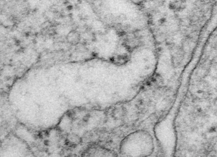
Pseudocolored electron micrograph depicting what I think is 4 separate actual protein transcription events within an RER profile — see as four ribosomes on the RER membrane and the resulting 4 strands of protein within the lumen of the RER. The RER cisternal profile is pseudocolored cyan, the ribosomes (4 of them in particular) are blue, the proteins being synthesized, 4 of them, from adjacent ribosomes are in red, and the extracellular space beside the type II alveolar cell (from a ferret) is brownish, and the type II cell cytoplasm itself is a light violet color. This was an accidental find while perusing hundreds and hundreds of type II cell electron micrographs from a half dozen species of mammal.

This particular micrograph was not taken FOR this purpose, but it illustrates a point in time frozen by fixative and plastic three decades ago.

9777_34065_RER_ribos_prot_color

Text book illustration electron micrograph: RER, ribosomes, protein production

This electron micrograph just made me chuckle because for whatever reason the fixation of this particular profile of RER just had four cute little ribosomes lined up and stringing out from them into the lumen of this profile of RER were four strands of protein, even to the point of having a little kind of fat area at the leading end….

Here is a contrast enhanced (I did use the burn tool in photoshop on the ribosomes and the protein) portion of that ferret type II alveolar cell, and below that is the area from which I cropped the profile of RER.  Magnification and enlargement can be approximated from the ribosomes… which are @ 25 nm in diameter.  Below that is the original micrograph, not retouched.

9777_31065_ferret_ribosomes_protein_inset9777_31065_ferret_ribosomes_protein9777_31065_ferret_ribosomes_protein_untouched