Daily Archives: June 22, 2016

Ferret type II alveolar cell RER showing parallel bands in organization of intra-cisternal protein

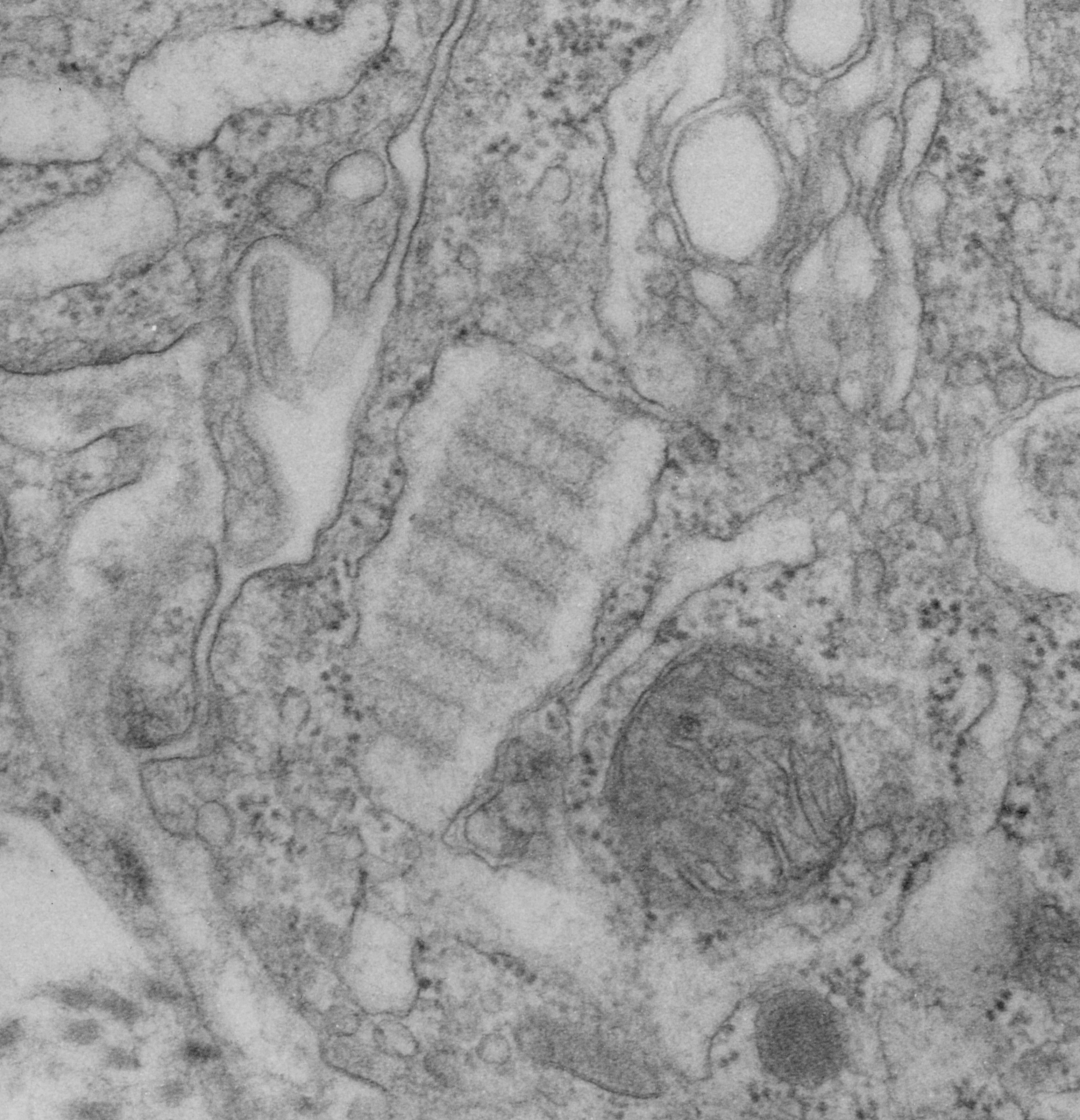
How awesome is this particular view of an intracisternal protein in a ferret type II alveolar cell. The bands are slightly tangential and so only the most electron dense banding is seen, however, the interesting thing here is that on the periphery there is not really any banding, but it looks as if the banding occurs centrally in the granule (aka within the profile of the RER).  Also, in the upper left of this electron micrograph (in an adjacent type II cell) examine closely the profile of RER  which actually can be perceived as ribosomes on the membrane surface and the trails of protein hanging off in the RER lumen.  Really a classic text-book presentation in “real life” so to speak, though we all know that TEM captures only a “nanosecond in time”, and all proteins are “fixed” and so distorted. Nevertheless, this may be one of those opportune views.

The protein (which I think is SP-A) is central in the micrograph, the banding pattern is at about a 35 degree angle.  Ribosomes are present on right and left borders for the most part, smooth ER is in the upper right corner and there is a tiny portion of a lamellar body off on the right side.  A portion of intercellular space with a couple of plasmalemmal folds crosses from top center to bottom left. There are a few other profiles of RER without SP-A? banding as well.

9777_31065_ferret_crop

Cat: periperhal lung type II cell

Lung tissue harvested from a sham treated control from another investigators experiment (thereby saving time – money – animals) here is a piece of a type II cell viewed with the electron microscope. It has my markings (for morphometry, back in 1982) and a marker arrow pointing to an area of RER which was the closest thing i could find to an organized protein in the RER (as a continuation of the search for species which may have SP-A organization similar to that found in guinea pig and dog and ferret.  In cat, though the total number of electron micrographs I have saved is not that great (a dozen or so) therefore the absence of such a layered intracisternal protein is not to be assumed).  The ribosomes studding the RER membrane provide an estimate of size (each being approximately 25 nm in diameter – a general number for all species and conditions…. (generalities are not always good for much, but an approximate size is OK here).

7296_24554_cat_3_lung

Type II alveolar pneumocyte of an owl monkey: electron micrograph

Still going through all the transmission electron micrographs of alveolar type II cells from 40 years of photography. Here is one print from an owl monkey (no treatment) I pseudocolored with yellow-brown for the nucleus, red for the nuclear pores, blue for the lamellar bodies, cyan (green) for the cytoplasm.  Just for the record, i did not find any protein structures within the RER which might be construed as intrcisternal layered RER inclusions that one wants to see if one is looking for SP-A.

owl_monkey_type_II_cell_7132