Daily Archives: June 1, 2016

Concentric organization of protein layered in an RER profile presumed to be surfactant protein A

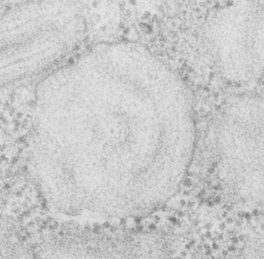
Guinea pig, aged, routine electron microscopy, type II alveolar cell, in particular the RER and a highly organized protein contained: possibly surfactant protein A.

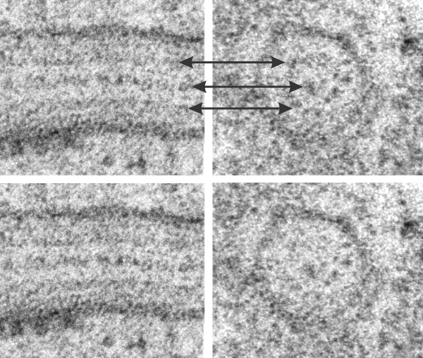
This micrograph is one of the few that is concentric and shows banding at the same time, at least that I have encountered.  The center period looks like the others which are only a single set of bands, i.e. a single central dark area, a lighter band, and then the outer band (which would include the CDR part of the protein octadecamers and the RER membrane.

It has ribosomes for size comparison, at 20-30 nm in diameter, and as can be seen from the longitudinal profile of RER in the upper left corner where the banding is more obvious, even in the concentric arrangement, the spacing of a single complete period is about 100 nm. I evened out the exposure of this micrograph, nothing else.

round_layered_ICB_gp

Longitudinal and cross sections of single period of SP-A? protein in guinea pig alveolar type II cell

From the same micrograph here are two tiny sections of the larger image which represent a well demarcated cross section (on the right) and a longitudinal section (on the left) where the organization of this protein is parallel to the RER membrane.  The banding in each is evident, however the bottom pair of images is “as is” and the top pair i have used the “burn” tool in photoshop to highlight where in the cross section the bands that match those in the longitudinal section batch up, and have added arrows as well.  One arrow to the central dot (which corresponds to the inner more dense band, presumably where the carbohydrate recognition domains in the trimer (or octadecamer) lie, and to the lighter band which may correspond to the N terminal regions.  It is my opinion that the N terminal groupings of the octadecamer come together in the highly periodic, but lighter bands, mirrored in the longitudinal section.

It is not clear how the inner CRDs fit into the central dot of the cross section…  particularly since the cross section is about 100 nm in diameter.  Picture on left has three or four ribosomes to compare size  (somewhere between 20-30 nm diameter) picture on right has one ribosome to compare.  Both are identical magnifications and imaging treatments.

cross_longitudinal_single_period_gp