Daily Archives: January 20, 2017

Single period alveolar type II cell granule: end-on

Electron micrograph of an alveolar type Ii cell which is overrun with surfactant protein granules and an isolated part of a larger granule shows this profile which is so symmetrical as to look like the spokes of a wheel. Micrograph on left is unretouched, micrograph on the right is burned in the areas of periodicity of the molecules of surfactant protein, and dodged in the areas between, just slightly to highlight what I saw.

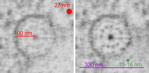
The purpose of the study is still to determine which of the surfactant proteins is responsible for forming the granules. So here is such a single period of a granule which has a ribosome marked for approximate size (red dot=27 nm) red bar=100 nm superimposed on an electron micrograph (unretouched). Right hand image has emphasized image, and a circumference of the round less-dense inner band of the period (central dense dot represents the “outer dense band” seen in linear profiles. The inner dots have been counted in linear granules at closer to 5-6 per 100 nm but here there are 11 or 12 dots of the less dense central band under the circumference, which is about 330 nm, or a little less, and in this photo the dots measure about 15-17 nm (consistent with previous measurements, then are spaced at about 25 nm apart. Not everything lines up exactly with previous posts on this subject in the linear profiles, but clearly they are analogous and match pretty well.  The spoke appearance likely corresponds to the vertical lineup of molecules seen in granules that have a linear arrangement. (just as an aside, there is something spooky about the fact that I have found several times now that the space between these molecules in the central less dense band are spaced apart the same distance as a ribosome…. that is just uncanny, and I bet it will end up meaning something about protein synthesis in general–just a heads up).  see this post and this post

6758_17082_gpig_301_single_circle

Where does the alveolar type II cell granule exit the cell?

Where does the alveolar type II cell granule exit the cell? This is problematic, as I have not actually seen a granule exit the cell at the apical plasma lemma or anywhere, for that matter, on the lateral or basal plasmalemma. This phot is probably a glimpse of the closest encounter between a granule and the apical membrane that I have noticed, to date. This is still not definitive. Wavy lines=apical membrane, arrow points to the cytoplasm between the alveolar space and the granule, which is smeared by the tangential orientation. Close, but not proof.  guinea pig, probably animal 301.

guess_guinea_pig_301

Desmosomal – mitochondrial associations

For at least three decades I have seen these associations, some more clearly defined, others not so clearly defined (like this example) of mitochondria that show cytoplasmic filaments adjacent, and in very close proximity to the plasmalemma, and to that of an adjacent cell.

This particular mitochondrion (M) is in an alveolar type II cell from a guinea pig lung and the desmosome is very indistinct but still clearly visible and the cell adjacent is very likely an alveolar type I cell due to its thinness.

I reported these in an obscure journal sometime in the 1980s, desmosome-mitochondria-associations as a cursory report, and then never did anything with it.  (aside: I do remember contacting some pathologist for information and to see if he had seen anything like this reported before…three months later, before i finished this paper, i saw his publication on this topic…. i learned a valuable lesson from this, not a negative one, but if one has to protect one’s ideas, then that must show that one doesn’t have that many.. i just accepted this theft).  So now I am returning to many micrographs (about 20k) and gradually finding examples of all the desomosomal – mitochondrial associations like this, to assess the frequency, the reason, and just add some general knowledge on the complex.

Red dot=approximate diameter of a ribosome to translate into 27 nm as a scale marker, M=mitochondrion, the very faint dotted circle has within the increased density between the mitochondrion and the very faint desmosome.  Photo has not been enhanced to show the desmosomal-mitochondrial connection.  10236 M8060 guinea pig alveolar type II cells.

10236_M8060_gpig_type_II_des-mit

Facebook: an online mob

People! Please understand the mechanics of facebook if you use it. It is an instrument, a medium, tool, just like dynamite and antibiotics.  You can use it for good and for evil. You can unknowingly use it for good, and unknowingly use it for evil.

Facebook is like a child, it craves attention. You can reward good behavior (like and friend people who make a positive contribution), you can reward bad behavior (unfriend the post, report it), whichever one you choose, YOU are an accomplice.  If you repost terror, abuse, destruction, cruelty, then you are an accomplice to those acts because you give them voice, and validity. If you punish a child, you still reward them… the behaviors become set.  If you repost goodness, honest reporting, facts (not just fake facts), then you are helping humanity.

Legally, if you stand by a friend while he/she shoots and kills someone, you are an accomplice, you can be tried and convicted. if you watch on facebook, cruelty and abuse, then you become an accomplice by rewarding inhumanity. (Too bad you can’t be held accountable)(yet).

Be responsible.  It is a mob of thoughtless and unconscious reposting and fake news that has the potential to engender much evil and hatred.  Facebook is no different than a stoning mob or a lynch mob for evil… it is mob behavior, fueled by anxiety, and depravity.  It can be a mob for good, a march and a crusade, fueled by love and by caring and by the desire to make the world a better place.