Monthly Archives: February 2017

“We are all much happier” said no one with a brain

I makes me wonder about someone’s mental capacity when they stand on a platform and say that economic uptic is their doing after 30 days in office. Worse yet to ascribe to everyone a “very much more positive outlook”…. that is not what i hear, nor see, nor feel.

AND…  for one who has been the ringleader in uprisings against other nations, races, religions,  one wonders why he says things like “We’ve got to keep our country safe,” Trump told the crowd. “You look at what’s happening in Germany. You look at what’s happening last night in Sweden. Sweden! Who would believe this?”  when he has initiated protests, riots, incidents right here in the US.   His anger is part of the new MOB-AMERICAN

Bet you can’t eat just one!!!

Bet you can’t eat just one.  Who perpetrates that kind of debilitating and for-profit advertising.  This has been around for half a century… bad on you Frito-Lay.

One of the reasons this tends to be a self fulfilling prophecy is that the brain just craves (and craves more) salt fat and sugar — the “satiety quick fix”.I bet you can’t eat just one carrot, or broccoli floret, or radish, said no one ever.  Why? because over the millenia of environment – good health interactions, we have grown up with the good things to eat.  They don’t turn us into mindless food addicts.

Who does this?

Only unamerican, self loathing, highly insecure, self righteous persons with low functioning frontal lobes, tweet out their anger to 34 million (and counting) people in all caps.   “SEE YOU IN COURT”  Truly the act of a undisciplined 2 year old mind.

Saving grace and justice: there will be thousands of nonsensical tweets documented for all eternity to laugh at. Hopefully we will recover from this style of governing.

Fuzzy ball vs ribosome

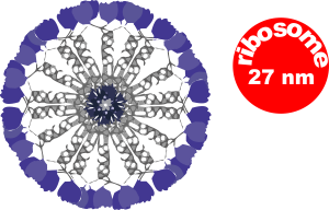
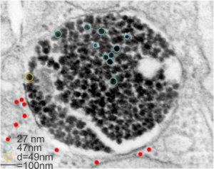
Here is a comparison, a diagram of what might be an SP-A fuzzy ball at something like 47-50 nm, and a nearby ribosome @27 or so nm.  The image on left, shows inset box from the granule in this alveolar type II cell from which the inset is derived.  My diagram isn’t exact, but it might be close.  These images are from guinea pig (now famous) 301.

Verge of a Dream: Illustrate my life

Someday. Not that it would
be hung, no one else could
illustrate my life.
With no care for it at all.
To pull the struggling
you give and give
what you have to
free someone from their
mud. Without repayment
only your attachments
are worth a fight.
There is an argument
you could write about
my life, as though anything
might change or matter. Like
terracotta, it starts from
dust and so it is done.
Your life much more
to say, without the
tarnish, will slip as
too many do, unappropriated.
Though with only
two sides and given
away, your gifts were,
to others, seeming to have
been too precious while
of no meaning, or these
coins were probably
much less to you. RLB 2 7 17

Are these fuzzy balls surfactant protein A or SP-D?

There is one guinea pig in this group of animals that was completely different, in the sense that all the surfactant granules  within alveolar type II cells in this animal are just overpowering all other cytoplasmic elements. This was a control animal which makes for interesting speculation, maybe even an oblique reference to the fact that this structure is a response to environmental influence (in this case probably a massive pulmonary infection or immune disorder. At least there was no experimental reason for alveolar type II cells to display so many granules.

Posted previously are images of an interesting variation on the granules, those which, in between the 100 nm layering pattern that is most prominent, some fuzzy rounded ball-like groups of protein exist.  A previous post that mentions these structures, in that case they were given a dimension of 50-60 nm… here I measured them more like 49 nm… so basically the same.  I was wondering at that time if they were surfactant protein A or not.  I am wondering now if they might be surfactant protein D.  Well, still leaning toward SP-A, in a configuration that would be two molecules “deep” or half the width of the banding seen in the granules.

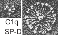
I did find this reference from Crouch et al, 1994 which had rat SP-D images that were awesome, however the dimensions don’t quite fit… and they give 46 nm as the size for a single arm of the SP-D molecule, and that is exactly half of what I measure as the full diameter of one of these dense structures diameter (47 nm). They calibrated their microscope well, I am not arguing with their final dimensions. From their paper are two screen shots C1q (left) and SP-D (right). The magnification is not given in a micron marker bar, but C1q is just a little smaller than SP-A, there fore if the relative magnifications are the same in these two, then that makes SP-D about twice as big as SP-A would be so maybe 50 microns which sort of contradicts the size given.  If C1q has a spread of the CRD close to SP-A (which is 20-20 nm) and picture on left is 20-30 nm diameter, then it appears that the SP-D here is about 2.5 times larger than SP-A would be and that is more like 70 nm  or even 100 nm, and a previous post on this blog calculated the round SP-D at 100 nm.  I am opting for the fuzzy balls in the granules of alveolar type II cells in this study to be SP-A.  Per diagram.

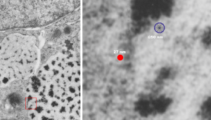
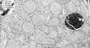
The image below (9124_17082_gpig_301_alveolar type II cell with granules, lamellar body (upper left under the legend) and a fuzzy all inclusion (in the RER i believe).  I saw these fuzzy balls embedded within granules but this is the only time I saw a cluster of them as a separate granule. Image below show the fuzzy balls clustered on the right, the figure below that shows relative sizes of ribosomes (red dots of 27 nm) and some examples of the diameter of the fuzzy balls at something around 47 nm and an optimally sectioned fuzzy ball (greatest diameter) of almost 50 nm.


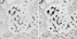
These fuzzy balls can occur, per above comments, within the granules which are otherwise typically without these spherical densities. micrograph 9864_17085_gpig_301. The red dots on right are more or less equivalent to 27 nm diameter (ribosome size) and the red bar is therefore equivalent to 100 nm.  Because of the lack of prominent layering and the added width (red bar) for the periods, it is presumed that this represents a tangential cut, and the size of the electron dense round balls (blue circles in images on right) is something around 47 nm (which is similar to estimates in other micrographs based on the size of a ribosome.  So these fuzzy balls can be seen both as separate membrane bound entities or found between the period layers of granules.  Unretouched photo on the left, photo on the right has the end-on single circular profiles highlighted…. looking like bulls-eyes.


Finding an alternative to alternative facts

From my son:

you’re using a term coined by the corporate media.  “alternative facts”
is an oxymoron. it’s like “conspiracy theory,” a term invented by
propagandists to discredit the rising alternative media… and by
alternative media, i just mean actual journalists who do research and
cite sources.  the corporate media doesn’t do journalism any more.

i love how their whole “fake news” meme backfired because everyone knows
the corporate media is the real fake news- and their falling numbers
reflect that.

the first step to not being brainwashed is to completely stop consuming
corporate media cnn,msn,abc,fox,cbs,disney,npr etc. all garbage, now
owned by just 6 companies (except npr of course which is foundation and
government funded, also lies).

no platform is perfect but if you use a variety of alternative sources
you will be much more informed.

below is a list of organizations who are replacing the dying corporate
dinosaur media. no single one of them is perfect of course, but they are
actually trying to do journalism instead of misinform and deceive.

“from me — I did find a website that i liked best of all…. justfacts.com… it gets point… no obscenities, violent speech, calls to riot, just lots and lots of graphs that show me trends and data. awesome”

jonrappoport.wordpress.com
infowars.com (questionable lately but in the past has done good work)
breitbart.com
wearechange.org
zerohedge.com
ae911truth.org
markdice.com
whatreallyhappened.com
therebel.media
ronpaullibertyreport.com
naturalnews.com
oathkeepers.org
thefreethoughtproject.com
michaeltsarion.com

social media platforms devoted to free speech:

gab.ai
voat.co
steemit.com

do not underestimate the value of this list.  there are a lot of posers
out there pretending to be alternative media to keep you from finding
the good stuff.  i’ve already sifted through the giant pile of crap as
best i can and this is the short list of the real stuff- those who have
a proven track record of accurate reporting and citing sources.

What do fixation and staining do

Methods of fixation and staining allow ultrastructural components of each cell type to be preserved and resolved differently. Glutaraldehyde based, non-coagulant-fixatives encourage direct cross-links with amines and other nucleophiles, most notably lysine and arginine, and cause deformation of the alpha-helix structures in proteins. Paraformaldehyde cross-links basic amino acid lysine, as well. Osmium tetroxide, in these studies was always used as a secondary fixative, except for beagle dog, where it was used as a primary fixative, helps to preserve lipids, and cross-links at unsaturated sites in membrane lipids and nucleophiles like amino and sulfhydryl groups. Osmium tetroxide enhances overall electron density of the specimen, adding contrast. Contrast is further enhanced by positive staining with heavy metal salts (uranyl acetate, enhancing lipids, nucleoproteins and proteins) and lead citrate (enhancing the contrast of proteins, membranes and glycogen) (Frankl, 2015; wikipedia). These all fit well with the type of layering (electron dense banding) found in alveolar type II cell granules.

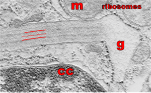
So what electron density in the structure of the alveolar type II cell reveal about the molecular structure of the layered protein. Well, surfactant protein A (and D also) has helical regions, probably hopefully the less dense central layers have a lot of lysine and sulfhydryl groups. This might fit well with surfactant protein A patterning.  Three red lines = 2 outer dense layers and less dense central layer; m=mitochondrion; ribosomes are the black dots (nucleoprotein staining with uranyl acetate), especially situated on the electron lucent racket end of the granule; cc= condensed nuclear chromatin (nucleoprotein staining with uranyl acetate), in the rounded portion of nucleus in the lower left corner of the micrograph. This image was shadowed with an exact replica, 68% transparency, and a few pixels to right and lower than bottom image. one of my favorite electron micrographs of these alveolar type II cell granules .