Monthly Archives: July 2017

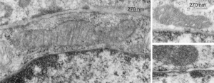
Several examples of mitochondria – nuclear pore interactions

These nice interactions between nuclear pore outer filaments and the outer membrane of the mitochondrion have intrigued me for at least 3 decades.  They first came to my attention when looking at liver, hepatocytes specifically, where they are frequent, and uncluttered by a lot of other cell machinery and organelles.  It seems pretty obvious that their tethering an energy source to fuel translocation of cytoplasmic proteins into the nucleus: “proteins (such as DNA polymerase and lamins), carbohydrates, signaling molecules and lipids moving into the nucleus” according to wikipedia. The author of the wikipedia post suggests that 1000 translocations per pore complex occur each second. Smaller molecules diffuse through at the peripheral portions of the pore and specific nuclear localization sequences are recognized by the nucleoporin proteins to assist larger molecules through the central part of the pore channel. That author also suggests that the pore transport does not require energy, so there must be some function for the mitochondria found associated with nuclear pore complexes (and sometimes appear to be just bent and dragged toward them). It is possible that assembly and disassembly of nuclear pores would require mitochondria. It has been suggested that three NUPs which do occur on the outer (cytoplasmic) filaments get phosphorylated. It is likely a testable hypothesis, which could be ferreted out at least a little in examining different cell types and relating that to mitotic index, or alternatively, from unperturbed to apoptotic state.  It is not that unusual to find three different areas of the same mitochondrion to be seen associated with outer nuclear pore filaments.

One mitochondria (on the left) shows two such close encounters.  These examples come from either alveolar type II cells or hepatocytes; grey dots 27 nm ribosome, bar=270 nm. It is interesting that the nuclear basket (filaments on the nucleoplasmic side of the nuclear pore complex) are not visible in any of these pore-mitochondrial associations, except maybe slightly in image on lower right.  In each micrograph, the mitochondrion is top side, a portion of the nucleus is below.
Patterns or artifacts: SER and outer mitochondrial membrane

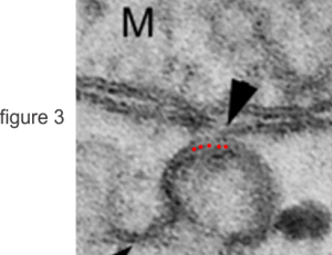
I just love it when I can see patterns in electron micrographs. I am not saying that there are not patterns in globs, and bumps and lines etc that appear as artifacts of fixation, of course they do, that is part of the nature of this particular process of inquiry. All methodologies have them, this one, that is the denaturing and folding and dis-alignment of molecules seen in aldehyde (or any) fixation, belongs to microscopy.  BUT WHEN i can see repetition in an orderly pattern as in these red dots, then i think, OK there is an underlying organization of the proteins (whether by fixation made lumpy or round or whatever), there is organization.  The authors that created this image have looked at mitochondrial-RER and SER interfaces and given reason to believe that there is a transport of Ca+ back and forth using SERCA 1 (sarco/endoplasmic reticulum Ca2+-ATPase 1s). They don’t mention the regular areas of protein denaturation here, but I think it would be highly possible that it relate to some aspect of the SER membrane proteins coming in close contact with outer mitochondrial membrane.  I added the red dots, just below actual dots in the micrograph,  highlighting the symmetry of spacing and size. Whether this relates to their fixation, or stain, their negative resolution or a structural organization on the SER i don’t know (the overall grain density of this micrograph is similar to the spot size), but it caught my attention. Their figure below is from —  Leopoldo de Meis , Luisa A. Ketzer, Rodrigo Madeiro da Costa, Ivone Rosa de Andrade, Marlene Benchimol . Fusion of the Endoplasmic Reticulum and Mitochondrial Outer Membrane in Rats Brown Adipose Tissue: Activation of Thermogenesis by Ca2+. PLOS One Published: March 2, 2010

Wait just a minute! is this amoeba lipid or intra-alveolar surfactant?

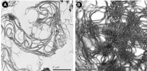
Wait just a minute! is this amoeba lipid or intra-alveolar surfactant? ha ha… I was googling liver toxicity and up came this article (from an image I selected to view the page) and i thought to myself… this could be intra-alveolar surfactant in the alveolar space (left) and tubular myelin in the alveolar space just organized in the center of the image on the right.  This just goes to show us that some principles and some patterns of molecules and physics are going to show up in surprising places.

The article itself was on 3D representations of some molecular organizations in cells, it was interesting as well and linked here.

mitochondria and SER: liver

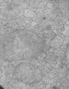
This micrograph has some interesting features for an ostensibly normal mouse liver: normal in that it is part of a study on Gclc loss, which causes early death  (specifics and article here) but this liver should not have had so many vesicles within vesicles as seen in the Gclc ko mice, and typically they are vesicles within vesicles without ribosomes.  here there are vesicles within the vesicles of RER …17941_73225_liver_Albwc_Gclcii_1mo_no_NAC

liver mitochondria and vesicle-within vesicles electron micrograph

Spit wad

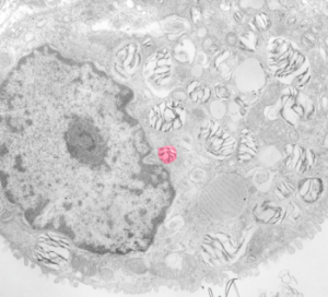
Sorry for being silly, but this alveolar type II cell just made me laugh, as I was dodging and burning to even up the micrograph I saw this cute little nuclear “face” and a little lamellar body spit-wad being sent out to the cytoplasm.alveolar type II cell electron micrograph joke