CAS names for the perfluorochemicals investigated for use as artificial blood (blood substitutes) are really important, as so many acronyms appear for the same chemicals. I have to say it has been a major headache just going through notes, and posts, and publications, to figure out the long names and CAS numbers for the acronyms used in the 1970s. I do thank the kind person at Fluorox labs (he might not want his name mentioned? i did not ask him) for some valuable info, and of course being a chemist, he suggested that I only use the CAS names (which i will try to do).
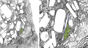
Two perfluorocarbons with at least one iodine atom, were made into emulsions and infused into mice in the 1973-1974 range by Leland Clark Jr (and the research faculty and support in his laboratory) one was called n-PF alkyl ethyl iodide (PFEI for short) and Fluorox Labs gives CAS 68188-12-5 to this compound and a second compound Perfluorodecyl iodide (IPFD) – presumably 1-iodoperfluorodecane) has the number CAS 423 621. I am really curious to see how these differe in vivo, since at the time, no one seemed to “have the time” to look over differences. There were 4 mice total, one mouse gave up several different tissues, from the others, only liver. Details below.
n-perfluoroalkyl ethyl iodide (PFEI) (CAS 68188-12-5)
Blocks 4759-4762 mouse 4, liver (one list says 4759,4760), liver
emulsion# 740313; PFEI 10% F68 5%, infused at 50cc/kg,
infused 3-13-1974 –sac date 1-14-75 (307 days)
perfluoro decyl iodide (IPFD) (CAS 423 621)
emulsion #730814, log#86 IPFD 10% F68 5%, 100cc/kg
2339-2342 mouse 1, liver (no osmium)
Blocks 2343-2346 mouse 1, liver
Blocks 2347-2350 mouse 1, kidney
Blocks 2351-2354 mouse 1, spleen
Blocks 2355-2358 mouse 2, liver (no osmium)
Blocks 2359-2364 mouse 2, liver
emulsion #730814, log#86 IPFD 10% F68 5% 10cc/kg
infused 8-14-73 –sac date 5-8-1974
Blocks 3774-3780 mouse 3, liver
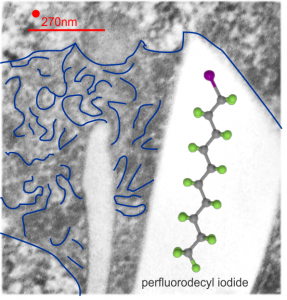
This is what I believe is the structure (inset within the large, presumed, crystalline footprint formed by IPFD; grey carbon atoms, green fluorine atoms and a fuchsia atom being iodine). Thanks to Fluoryx labs and Chemspider and wikipedia and of course google.
Two white rhomboid spaces where IPFD was once in these lysosomes are seen (vertical spaces–one large one quite thin), and the blue outline is the apparent limiting membrane of the lysosome (which encloses the crystalline IPFD), and the blue lines that wiggle within the lysosome show a sheet-like-periodicity that needs further investigation but is very clearly a pattern in most of the lysosomes with IPFD inclusions, and not really present in lysosomes from the other artificial blood substitutes that I have looked at with TEM (that would be a long list).
Red dot is ribosome to estimate size at 27nm, red line is micron marker derived from ribosome size. No measurements yet made on the longest crystal length, but it is going to be many microns for the largest, and the directionality has yet to be associated with any cytoplasmic elements.
