If I fail to try something that frightens me I will forever be a slave to it
Monthly Archives: January 2018
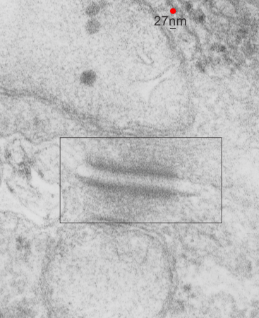
Rhesus monkey hepatocyte desmosomal mitochondrial tether
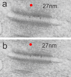
There are some ultrastructural characteristics of this desmosome which are interesting. Having a desmosome sectioned opportunely is hit and miss. This particular section has some structures that look to be the correct size of intermediate filaments that are trailing off the the upper right under the mitochondrion, and there are definite light-dark-and dotted portions of the desmosome (in and including the plasmalemma portion) which have a distinct vertical repeating pattern.
I squeezed the micrograph bottom to top to determine if this was just a shifting of the image during photography or whether these vertical striations would still be visible when i removed (in photoshop) that kind of vertical shift. The answer was that even then the spacing and the effect of the alternating light and dark areas in the cell membrane portion of the desmosome remained. Red dot=27nm as determined by ribosome size, and thus the alternating dark and light vertical areas in the section are about 10nm in width.
In addtion, there is a clear darkening of the outer mitochondrial membrane, and a slight darkening of the inner mitochondria membrane at the point of the desmosomal-mitochondrial tethering. One can suggest that there is a relationship between this patterning and the various protein families that comprise the full structure of the desmosome, intermediate filaments and mitochondrial connection.

Baboon hepatocytes and single desmosome
Control animal (neg 5936 block 8830 baboon, f, #23, right lobe of the liver, control bx, modified Karnovsky’s fix, 1% osmium tetroxide, Epon 812, (9 28 1977)). There are a lot of smooth vesicles in this sample and just a little bit of RER, three lipid droplets, and part of a bile canaliculus, possible the space of Disse in the very upper most right part of the image, mitochondria look pretty normal) I was looking for desmosomal-mitochondrial tethers and found this pretty nice desmosome and I pseudocolored the plasmalemma from the two adjacent hepatocytes present at the desmosome purple. There was a clear the “outer dense layer” of the desmosome (which i think is so unfortunately named “outer” and should have been named “inner dense layer” of the desmosomal complex). It had a particularly “loopy” appearance that I highlighted in orange. Top image is the original micrograph (two hepatocytes) and box is the area enlarged in the figure below with a desmosome).

