Category Archives: Layered intracisternal protein granules in mammalian lung

For 35 years I have thought about this protein organized so nicely in the type II cells of the lung of three mammalian species (guinea pig, ferret, mongrel dog) which I am sure is present in more species, and have not had the time to investigate it further. A peer review publication in the early 1980s was about as far as the study went, though published in an OK journal (Ultrastructure Research) I felt there was more to see than just what I could put down in those few pages. Life makes us make choices, and at this point I choose to play with this old problem. I am not professing anything except diligent “musings” here.

RER protein in a type II cell of a guinea pig

The stacks of RER cisternae in this particular type II cell don’t show the layering and patterning that other cisternae from these animals do, that is, they look more like ordinary RER cisternae (though clearly a little dilated, and clearly more stacked).  Still I think this is an overproduction of SP-A or some SP as other images from the same animal show the periodicity seen in intracisternal bodies.

The stacks of RER are reminiscent of the stacks of birbeck granules in the Langerhans cells where langrin is overproduced.  This is a paper by  J. Valladeau et al, Immunity 12: 77-81 (2000) where they transfected langerin cDNA into fibroblasts and got stacks of birbeck granules (which they show in an electron micrograph). While the image below is certainly NOT langerin, and NOT a transfection of any SP-A cDNA into alveolar cells, it is probably the ONLY image that i have found where the profiles or RER might mimic granules (RER) of other c-type lectins (like the birbeck granule) in terms of being segmented and stacked.

type_II_cell_RER_and_LBs_gp

Who has electron micrographs of c-type lectins in situ, in vivo

I am continuing to search for electron micrographs which have images in high resolution and magnification of the c-type lectins to compare with the intracisternal protein accumulation in type Ii cells of the lung of some species of mammal.  Upon thinking about the, comparing with the fine structure of langrin in the Langerhans cell, it occurred to me that I might have a similar structure in the microvillar cell of the olfactory epithelium.

The microvillar cell has not been a topic of much research, and it has no well established function. Because of the occurrence of about 10% of most epithelia in the body to be burried in the epithelial cells proper (10% in olfactory epithelium, back skin, colon epithelium, etc) that I have examined personally it made sense to go over that set of about 250 micrographs looking specifically for RER profiles that specifically had a periodicity or layering to the protein content.  Unfortunately, in all those microvillar cells examined, I did NOT find any organization to the protein contents of the RER profiles, but I did find the sparse ribosome studding on the profiles that I have seen in the type II cell of the lung.

Nothing conclusive, but just a little bit peculiar.  I do wish I had seen some indication that the RER of the microvillar cell had a “granule” like the birbeck granule… ha ha….  that would have given an instant functionality to the microvillar cell.  It might still be there… as not all RER protein contents show periodicity like the one in type II cells and Langerhans cells.  I have not found images actually for any other collectin that speak to layering, just the one I have examined and the birbeck granule in Langerhans cells.

Here is a quick composite of some profiles of RER in the microvillar cell of the olfactory epithelium (recall, that upper layers contain the olfactory bipolar cells, the sustentacular cells (supporting) and microvillar cells (at about 10% occurrence.  Profile of nucleus in the bottom sector of each image, perinuclear space and RER profiles are just above that.  RER profiles are quite electron lucent, and only on e of the pictures shows any contents (and to me those are too close in shape to ribosomes to be excited about).

I would love to see any electron micrographs of MBL or other c-type lectin protein overexpressed in any type of cell.

microvillar_cell_RER

Concentric tubular arrangement for surfactant protein in RER of type II cells

More images which pique the imagination about what is responsible for the highly organized protein in the RER of the type II alveolar cell in some mammals (in this case an aged guinea pig, control, from a previous study).  I have mentioned before that there are examples of wildly converging and blending and mixing of these proteins going from a perpendicular arrangement to parallel to the long axis of the RER profile and splitting off and curving and also concentric or semilunar.  This is a really nice resolution and relatively clear image of a tubular arrangement, cut in cross section, comprising a single “period” that is here seen as center dot, medium density ring and outer ring.  This short video is constructed from two images, the initial image is untouched, the fade in image i have used photoshop “burn” tool to highlight the area that looks to me like it is concentric.

I added a blue color to an overlay of the actual cisternal body on the left, and a brownish overlay to the nucleus at the bottom right corner.  Ribosomes are clearly found, and are especially prominent where the banding runs parallel to the RER, just above the tubular section.

Perinuclear space might have a trace of this protein in it, perpendicular to the direction of the nuclear membrane but it is quite indistinct,  and there is a perichromatin granule very near the nuclear pore seen on the left hand side of this profile of the nucleus.

video is available on YouTube:

Composite image of intracisternal body (blue, nucleus, brown)

concentric_blended

Surfactant protein D rotary shadowed fuzzy ball

Here is an awesome picture in a publication by the following individuals: Martin van Eijk, Chris H.A. van de Lest, Joseph J. Batenburg, Arie B. Vaandrager, Joseph Meschi, Kevan L. Hartshorn, Lambert M.G. van Golde, and Henk P. Haagsman “Porcine Surfactant Protein D Is N-glycosylated in its Carbohydrate Recognition Domain and Is Assembled into Differently Charged Oligomers”, American Journal of Respiratory Cell and Molecular Biology, Vol. 26, No. 6 (2002), pp. 739-747, which I have cropped and repositioned and added a micron marker to.

I am trying to sort out whether the banded protein in the RER of some guinea pig, ferret, and mongrel dog specimens that I have repeatedly observed could be surfactant protein A or perhaps surfactant protein D.  It seems to me that It is possible for the 100 nm periodicity with a central dark band to be surfactant protein D, but because of the relative abundance of SP-A in lung, it seems less likely, though not impossible that it is SP-D.  Another study by: Stefanie M Heinrich and Matthias Griese Assessment of Surfactant Protein A (SP-A) dependent agglutination BMC Pulmonary MedicineBMC series,  shows artificial organization of SP-A, which does the mirror aggregation as I have predicted in my images.  They used streptavidin beads in vitro to accomplish this, and while not showing images that I particularly “believed” the diagram they provided was similar to mine.

Upper image the cruciform tetramer, bottom image the SP-D fuzzy ball.

I think SP-D may not be the protein in the intracisternal RER (granules) profiles in guinea pig in my micrographs, because of the low electron density of the space between the CRDs, that similar area in the SP-A showing a greater relative electron density than seen in the fuzzy ball below.  This is also more reminiscent of the periodicity seen in the langrin/CD 207 protein in Langerhans cells (Birbeck granules), as you will recall that that linear density is electron pale and very electron dense, not more middle density like the intracisternal protein in guinea pig type II cells in these posts.

surfactant_D_rotary_shadowed_SP-D


Concentric organization of protein layered in an RER profile presumed to be surfactant protein A

Guinea pig, aged, routine electron microscopy, type II alveolar cell, in particular the RER and a highly organized protein contained: possibly surfactant protein A.

This micrograph is one of the few that is concentric and shows banding at the same time, at least that I have encountered.  The center period looks like the others which are only a single set of bands, i.e. a single central dark area, a lighter band, and then the outer band (which would include the CDR part of the protein octadecamers and the RER membrane.

It has ribosomes for size comparison, at 20-30 nm in diameter, and as can be seen from the longitudinal profile of RER in the upper left corner where the banding is more obvious, even in the concentric arrangement, the spacing of a single complete period is about 100 nm. I evened out the exposure of this micrograph, nothing else.

round_layered_ICB_gp

Longitudinal and cross sections of single period of SP-A? protein in guinea pig alveolar type II cell

From the same micrograph here are two tiny sections of the larger image which represent a well demarcated cross section (on the right) and a longitudinal section (on the left) where the organization of this protein is parallel to the RER membrane.  The banding in each is evident, however the bottom pair of images is “as is” and the top pair i have used the “burn” tool in photoshop to highlight where in the cross section the bands that match those in the longitudinal section batch up, and have added arrows as well.  One arrow to the central dot (which corresponds to the inner more dense band, presumably where the carbohydrate recognition domains in the trimer (or octadecamer) lie, and to the lighter band which may correspond to the N terminal regions.  It is my opinion that the N terminal groupings of the octadecamer come together in the highly periodic, but lighter bands, mirrored in the longitudinal section.

It is not clear how the inner CRDs fit into the central dot of the cross section…  particularly since the cross section is about 100 nm in diameter.  Picture on left has three or four ribosomes to compare size  (somewhere between 20-30 nm diameter) picture on right has one ribosome to compare.  Both are identical magnifications and imaging treatments.

cross_longitudinal_single_period_gp

Could these be surfactant protein D fuzzy balls?

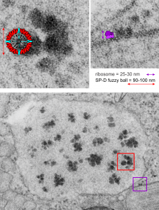
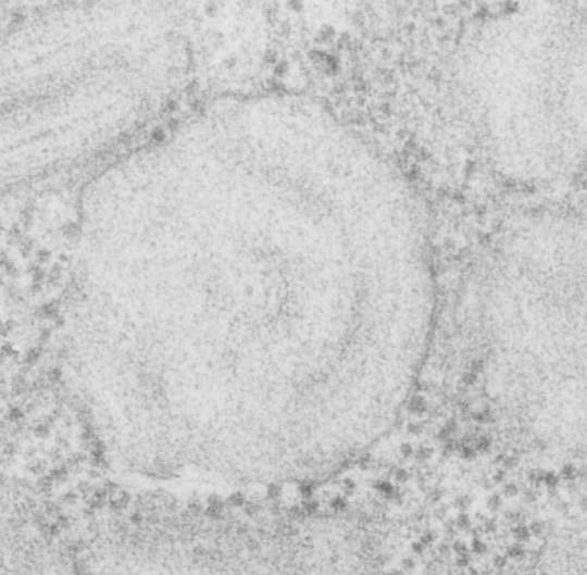
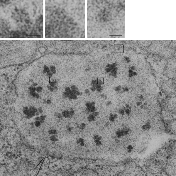
It is so fun to try to figure out whether the accumulations of layered protein in the RER in type II alveolar cells are, in fact, surfactant protein A.  These images have sat on my office shelf for 30 years.  As posted a few days ago, this particular electron micrograph has intracisternal bodies or granules or RER accumulation of a layered protein (call it what you want) that were common to guinea pig type II cells, but just one profile of RER had funny round “fuzzy balls” in it.  I only saw this once.  From the beginning it was easy to see that they were not at all like ribosomes, which one might quickly think could get stuck in the bands of such a wild production of a protein, but they are the wrong size, and have a  totally different texture appearance, so ribosomes are pretty much “out of the question” as a choice. The next easiest conclusion was that they were glycogen, also electron dense, but I am not sure why there would all of a sudden be glycogen in these protein (- whatever protein it turns out to be) accumulations, and besides, glycogen is more random in its clumped appearance and is bound within membranes (one can google pictures of glycogen in the liver and see the difference).  But I remembered reading about these wacky “fuzzy balls” probably composed of surfactant protein D (Patrick Waters et al, Chapter 6, in: Target Pattern Recognition in Innate Immunity, ed. Uday Kishore, Landes Bioscience and Springer Science+Business Media, 2009) and did some images tests to determine if these could be surfactant protein D.  Fuzzy balls are the cruciform surfactant D, becomes a multimer  – 4 crucifers in the same 100 nm (others say more like 90 nm) structures.s

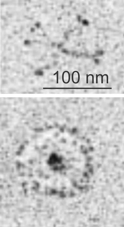
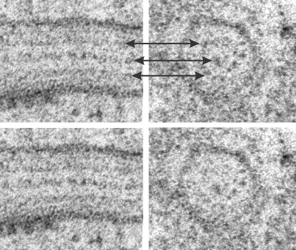
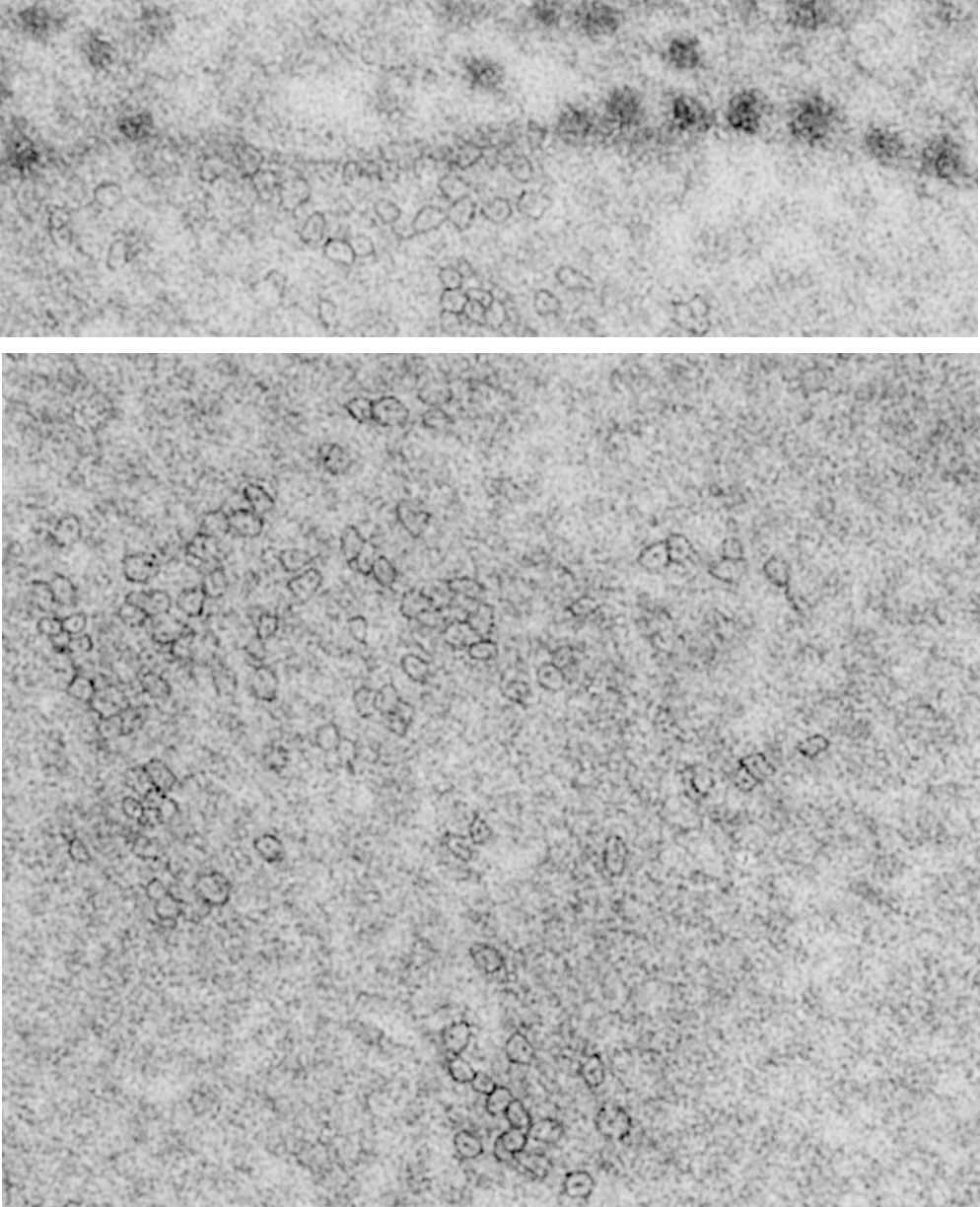
I just cropped out ribosomes from the identical electron micrograph as I cropped out a sample of these fuzzy round structures and compared their size, side by side.  You see that this doesn’t come out exactly right.  The proposed size for the surfactant protein D fuzzy balls is just a little bit too large, to fit nicely into the shape of what appears in my micrographs.  In addition, I was hoping that the only protein found in these intracisternal bodies was surfactant protein A, and not containing any other surfactant protein.  But the thought was intriguing.  I do not know of any reports that indicate surfactant protein A can form similar fuzzy balls, but it seems that it might be possible…. and the dimensions for a surfactant protein A fuzzy ball would be closer to the size estimated for these, per the adjacent ribosomes.

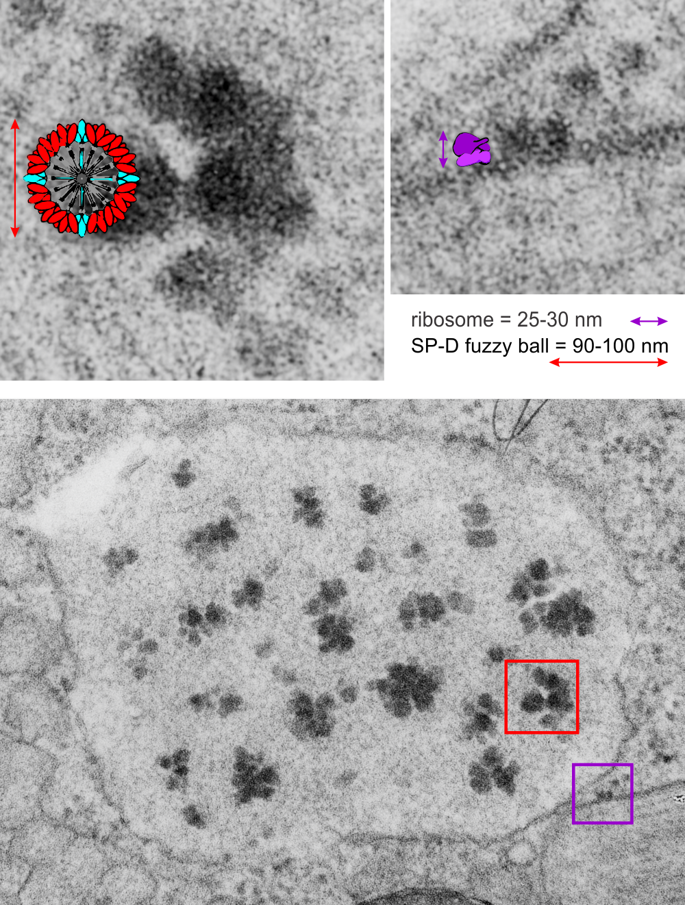
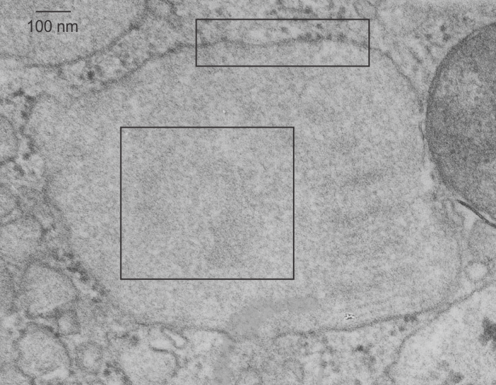
Anyway, check out the diagrams of a surfactant D fuzzy ball and a ribosome, matched with their nm markers, and the micrograph of the intracisternal body from which the fuzzy balls were derived.  If you have any interest, or information, I would love to hear about it.  Red rectangle on bottom image is the portion of the fuzzy balls used, while purple rectangle on the bottom image shows where ribosomes were used.

fuzzy_balls

Dark band of RER profile in type II alveolar cell of a guinea pig

Dark band of an intracisternal body (dark bands in a perpendicular section of these intracisternal bodies span about 100 nm, but when spread out over a tangential cut, can have many more nm from the next dark layer in these inclusions. (I am calling them inclusions for want of a better word, but are rather sites of over-production and layered organization of what I think is SP-A.  It provides an alternative view of the very orderly periodicity (3,5,7,9 – depending upon orientation and how thin the section is).  These two images are identical except that I have increased the contrast, and burned (photoshop) the order which I see in these tangential portions (which likely represent the carbohydrate recognition domains of SP-A.  I thought at first these hexagons were SP-A, then began to wonder if i could find similar structures “anywhere” in “any section” as an artifact of fixation, so went to several micrographs and scanned and examined them for randomly occurring small octagonal structures about 25 nm in diameter.  There are many, but then I used the “light” banded area just as a control to the dark banding in an RER profile though to contain large amounts of organized SP-A as an internal control, and to help rule out the possibility that the fixation turns all proteins into patterned molecules (which it probably does to some degree) but which would fool one into thinking it was SP-A.  So the bottom line is that I think the hexagonal structures (about 20-25 nm in diameter (see ribosomes nearby for comparisons in size at 20 nm).

tangential_guinea_pig_ICB_dark_band


Unusual RER profile with SP-A? protein plus other inclusions.

This particular profile of RER in a type II alveolar cell of a guinea pig has bothered me for at least 3.5 decades. The guinea pig is aged but otherwise untreated, i.e. wasn’t part of a experimental treatment protocol but was part of a study on toluene diisocyanate exposure.  This RER profile has very large cluster inclusions within the periodicity, which is very difficult to see in this particular profile because of the tangential sectioning.  I have no clue what this is, but it shows considerable substructure on enlargement of the micrograph (taken at 23,500x, with the V pole piece in an old Siemens 1A electron microscope, and the enlargement in the darkroom was 4x.  Scan was 3200 ppi.

Several areas which showed a terrific hexagonal substructure were cut and pasted, a marker for their relative nm bar scales given in each, based upon a repeat measure of local (the same micrograph) ribosomes at a pretty much standard 20 nm size.  Please help identify these (ha ha) (I do have comments turned off for obvious spam reasons) you will be able to contact me in other ways.

tangential_guinea_pig_ICB_odd_inclusions


Guinea pig alveolar type II cell: SP-A bouquets?

Plugging away at whether these hexagonal structures found within the RER protein accumulations in some alveolar type II cells which are found on tangential sectioning of what I have called “intracisternal bodies” are really SP-A bouquets which are splayed out with a center dot being the neck and collagen portions of that molecule.  I have taken a portion of one such intracisternal body which has a small portion which is ribosome studded highlighted (using the burn tool in photoshop) a few of the hexagonal arrangements which seem to have six or so densities on the perimeter, possibly the carbohydrate recognition domain trimers of the SP-A 18-mer.

Previously posted images of those found in ferret type II alveolar cells really looked to be larger, and that is problematic.  These hexagonal structures (pictured below) are from an aged, untreated, control, guinea pig  and the bouquets seem more in line with the size of a 20 nm ribosome, a string of which I have included for assisting with perception of relative sizes.  There is a large tangentially sectioned dark band (actually two dark bands) in the image below unaltered, enlarged with the ribosomes in which I have highlighted the hexagonal molecules (using ONLY THE BURN TOOL in photoshop) to accentuate what I see as patterning. The ultimate test of this would be to take an unrelated micrograph of an unrelated RER inclusion, at the same magnification and see if i could pick out as many hexagons per nm in it as I find in a tangential section of an intracisternal body ( ha ha–sort of pedestrian but it would be a good test).

So the upper micrograph gives you a distance marker (bar=100 nm) and the upper box gives you the area of ribosomes and a portion of the intracisternal protein structure which I presume to be SP-A. The lower box gives you the identical area of as is found in the bottom panel, which has two areas of slightly darker electron density marking the tangential portions which are the dark lines of the intracisternal body periodicity.  In both enlargements (boxes) I have highlighted just a few of the very many hexagons, which are hopefully the top down view of SP-A molecules.

For those of you who are curious, a mitochondrion is off to the right, and there are also portions of two large intracisternal bodies, and half a dozen or more single – periodicity intracisternal bodies as well.  In the central intracisternal body on the right hand side shows the 100 nm periodicity (which on a completely perpendicular section contains anywhere from 3 – 9 bands).

guinea_pig_ICB_SP-A_boxesguinea_pig_ICB_SP-A_highlights