The gastric epithelium was a research topic of several years ago, and for one submission of a manuscript for publication I also created a cover submission and submission to Bioscapes.
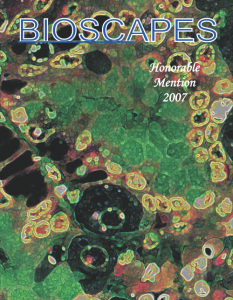
The gastric epithelium was a research topic of several years ago, and for one submission of a manuscript for publication I also created a cover submission and submission to Bioscapes.
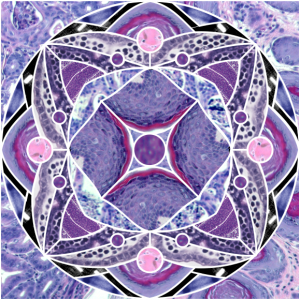
There are few things in life that can be separated distinctly from each other: science and art are some of those things. This diagram is a compilation of computer graphics, geometry and actual images of mouse back skin taken from a legitimate and productive research project. Who would have guessed that follicles and dermis could be so fun. Below is a cover submission to an online journal that provides help for graphic artists. 
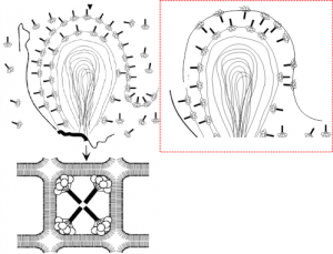
An article by Palaniyar N, Ikegami M, Korfhagen T, Whitsett J, McCormack FX. 2001 entitled Domains of surfactant protein A that affect protein oligomerization, lipid structure and surface tension, published in Comparative Biochemistry and Physiology 129:109-127, presents a diagram of how the 18-mer of SP-A might integrate itself into the outer lamellae of lamellar bodies in alveolar type II cells. I did notice an inconsistency, perhaps, in the diagrams. The bottom diagram (left bottom) shows the SP-A molecules with their N terminals all pointing inside, or towards each other in the center of these tubular myelin structures (the diagram is a 2D model of a 3D structure, hence “tubular”). In the granule found in alveolar type II cells which I have been trying to describe morphologically for several years has a layering pattern that suggests that the carbohydrate recognition domains are actually pointing “away” from each other. This is replicated in the diagram to the left. The inconsistency comes in how the SP-A molecules are represented in the top portion of the diagrams, which I duplicated in part, and re-oriented the SP-A molecules to match the tubular myelin arrangement shown in their figure (bottom left). To me this makes more sense, and it also is consistent with the layering of the alveolar type II cell granule in guinea pit, ferret, and dog, and in addition, with the outward orientation proposed for the Birbeck granule (the C-type lectin, langerin). Comments are welcome. My edits to their diagram are in the red dotted inset to right.
This electron micrograph is from a cat lung, and is an alveolar type II cell. There are some areas of dilated RER which seem to fit the description of a surfactant protein A type organized granule. In particular this banding, which is admittedly faint (not section chatter as just above it there is the membrane of a mitochondrion which is pretty much in line with the middle band of this granule which might make one think the banding is chatter, but the dense line does not extent further into the micrograph past the rounded edge of the mitochonrion, therefore is not likely to be chatter.
I think it is the best example of layering (oligomerization of the protein) within the RER that I have of the few electron micrographs of cat lung that I have.
A lamellar body is within the image on the left of the nucleus in the lower magnification view. The enlarged portion is demarked with a white box (in the left hand image). Judge size by using the two ribosomes on the upper membrane of the perinuclear area that has the layering (layers are pointed out with black arrows). Ribosomes would be about 27 nm in diameter, making the banding about four ribosomes wide…. or more or less equivalent to the 100 nm layering predicted to be SP-A. Yep, circumstantial, but quite likely.
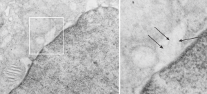 Cat number 3, alveolar type II cell electron micrograph, neg 7268 block 24564, original magnification 5000 x, enlarged 4x.