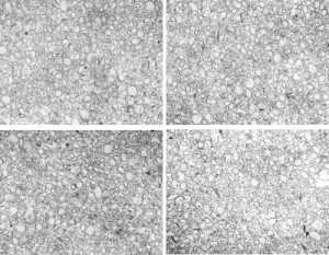
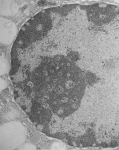
Really nice electron micrograph of liver nucleus and enormous nucleolus, lots of nuclear pores and perichromatin granules and condensed chromatin. 16029_65718_no_NTBC_14CoS/14CoS ko
 Abstract from a paper by Deiter et al, follows: Whereas ch/ch wild-type mice and ch/14CoS heterozygotes are viable, 14CoS/14CoS mice homozygous for a 3800 kb deletion on chromosome 7 die during the first day postpartum. Death is caused by disruption of the fumarylacetoacetate hydrolase (Fah) gene; absence of FAH, final enzyme in the tyrosine catabolism pathway, leads to accumulation of reactive electrophilic intermediates. In this study, we kept 14CoS/14CoS mice alive for 60 d with oral 2-(2-nitro-4-trifluoromethyl-benzyol)-1,3-cyclohexanedione (NTBC), an inhibitor of p-hydroxyphenylpyruvate dioxygenase, second enzyme in the tyrosine catabolic pathway. The 70% of NTBC-treated 14CoS/14CoS mice that survived 60 d showed poor growth and developed corneal opacities, compared with ch/14CoS littermates; NTBC-rescued Fah(-/-) knockout mice did not show growth retardation or ocular toxicity. NTBC-rescued 14CoS/14CoS mice also exhibited a striking oxidative stress response in liver and kidney, as measured by lower GSH levels and mRNA induction of four genes: glutamate cysteine ligase catalytic (Gclc) and modifier (Gclm) subunits, NAD(P)H:quinone oxidoreductase (Nqo1), and heme oxygenase-1 (Hmox1). Withdrawal of NTBC for 24-48 h from rescued adult 14CoS/14CoS mice resulted in severe apoptosis of the liver, detected histologically and by cytochrome c release from the mitochondria, increased caspase 3-like activity, and further decreases in GSH content. In kidney, proximal tubular epithelial cells were abnormal. Human hereditary tyrosinemia type I (HT1), caused by mutations in the FAH gene, is an autosomal recessive disorder in which the patient usually dies of liver fibrosis and cirrhosis during early childhood; NTBC treatment is known to prolong HT1 children’s lives-although liver fibrosis, cirrhosis, hepatocarcinoma, and corneal opacities sometimes occur. The mouse data in the present study are consistent with the possibility that endogenous oxidative stress-induced apoptosis may be the underlying cause of liver pathology seen in NTBC-treated HT1 patients.