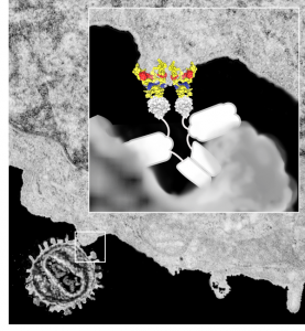
I made this diagram for someone a decade ago, whose name (LS) means nothing to me and the names of the protein complexes escape me. It was a cover submission for an article submitted to PNAS at some point and didn’t make the cut, and I am sure LS never paid me. It has some interesting information in it anyway so I am posting it as an illustration. This version is considerably modified so I am not breaking any copyrights…besides it was my illustration in the first place (LOL).
Monthly Archives: May 2017
Great statistics, much more interesting than politics
Here are some fun numbers from the NIH website (reduced to a skeleton, LOL)
- 3.2 billion base pairs for DNA
- 20,000 genes make 100,000 proteins
- DNA unwound in one cell placed end to end = 67 billion miles
- DNA from any two people are 99.9% identical
- One third of our genes are managed by microRNAs
- 98% of our DNA is not understood at this point
Bilateral nuclear symmetry
These images (taken from salivary polytene nuclei electron micrographs from a publication by Carla Ritagliati, et al, show me that there is symmetry in the nucleus regardless of what stage of either replication, growth, apoptosis, it is in. And while their publication has to do with totally different topics, i was struck by the duplicate nuclear domains within their images. Because electron microscopy is 2D by inspection of single micrographs of course, this might get translated into a radial symmetry so easily. Would that I had the equipment and time and funding to figure some of this out. This is from their figure 7 just cut and cropped the images which were obviously bilaterally symmetrical.
I can — I can’t
Where in the brain is the group of cells that tell me “I can” or “I can’t”?
How long before those cells send off the “I can” vs the “I can’t” signals?
What is a good way to maintain the “I can” attitude when sometimes it is impossible to to do what needs to be done until later
Everything we do impacts our brain — and in googling this i found an amazing (but I should have thought it existed) site, THE HUMAN CONNECTOME, go to their “publications” tab and read some of the interesting abstracts.
I think in a quick reading of some links about brain and body dualism, i would have to accept that it is NOT dualistic at all, but a unit, ha ha, i would avoid labeling it as Unitarianism.
And a great quick video on what the human connectome is all about is HERE, awesome and really amazingly complex. and an elementary explanation of their process for building 3 dimensional maps of tiny portions of brain (mouse i think) is in this video... I do have a question and that is how they get those slices to pass into a vacuum on a conveyor belt.. ha ha. I suspect some of the important steps have been left out. Of course i love this concept because I am an electron microscopist…. and have wanted 3D techniques all my research life…. at this point 40 years past when I could have gotten into this, I am loving that it is being used.
Bottom line is that at some point, some lucky person is going to know exactly where in their brain the impetus for “closing, or I can’t” vs “opening, or I can” is wired into your heads.
Defining the components of the interchromatin granule clusters
I have not found a good definition of interchromatin granule clusters. Beginning concerns are: 1) the clusters have non membrane bounds, this presents a perfect opportunity for misconceptions. Here are some predictions:
- inerchromatin granule clusters behave like lipid droplets (begin perhaps irregularly shaped but finally ends up roundish.
- Variations in the granularity of interchromatin granule clusters, coarse, fine, symmetrically placed, numerous and also scarce, large and small.
- Numerous protein-protein- protein-RNA interactions
- Abundance of different proteins, at various times, some of which can form oligomers such as SPOP (which can make 25-mers) and the degree of oligomerization may depend upon the concentration of protein in the interchromatin granule cluster.
- The cluster itself has boundaries that are clearly demarkated regardless of the number and size of structures with those boundaries.
- SPOP dimers help organize higher order structures within the interchromatin granules clusters and other places
Components of the interchromatin granule clusters are ??? and do they change during apoptosis
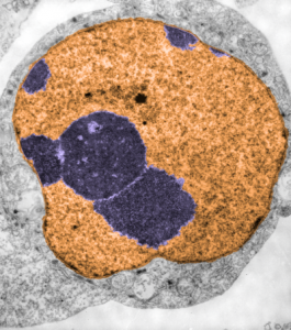
 While interchromatin granule clusters were thought to be the “the same structure” as nuclear speckles, i wonder if that opinion was justified at the time, I am not about to make that leap. I need to sort this out, since with plastic sections stained with toluidine blue, the interchromatin granules really were not “lucent or unstained” areas, but a light tan color, if i remember right, and with TEM they were actually more closely punctate than usual euchromatic nucleoplasm. So the connection needs to be further varified…and this might be why there are mixed notations and confusion and disparate explanations as to what the speckles and interchromatin granule morphologies identify. Also, what I have seen referred to as interchromatin granule clusters have two (at least) maybe three or four sizes of granular elements depending upon metabolic or functional state of the nucleus is (as in G,S and also stages of apoptosis, maybe necrosis as well. (Of course all the (11 so far) listed processes of cell death and cellular-self destruction could be listed here but I just subscribe to the main ones, necrosis and apoptosis for now.) This cell has nucleolus dark blue, nucleus golden, cell cytoplasm greyscale, interchromatin granule cluster of pink pink, and white box shows area enlarged in picture below. The larger (one quite large) and four or so smaller clusters can certainly point to some metabolic phenomenon which is not really occurring in a non-cultured-non-exposed cell.
The purpose here is to comment on the various sizes and shapes of such densities within the interchromatin granule clusters, and to examine transmission electron micrographs of so many cell-death projects to see whether concistent patterns interchromatin granule clusters have components change in size, position, density, and shape depending on which processes are occurring within the nucleus.
In particular, when A549 cells were examined after knockdown of an antiapoptotic gene (using si67), then the interchromatin granule clusters contained large aggregated densities (large in comparison to the smaller entities in untreated cells). Here I have localized these in a single cell (transmission electron micrograph of the area bounded by the white box above), just to point out what is observed. Relevance is yet to be determined for these larger than usual areas of of conspicuous density within interchromatin granule clusters. The curved arrow points to bar-like organization of a filament or fibrilar area (sometimes this ribbon type linear organization is seen in cajal bodies) and the width of these bar-like fibrilar structures would be something around 30-40 nm, if compared to the size of a ribosome (red dot in the center of the text that says 500 nm). The large structure (electron dense) beneath the interchromatin granule is the top part of the nucleolus seen in the image above. neg 18272 block 78932 A549 cell si67 knock down of antiapoptotic gene in vitro.  )
)
My kind of science art: Starry Night by Alex Parker
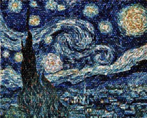 Wonderful images exist in science, I can attest to this having taken thousands and thousands of electron micrographs of the intricacies of the myriad cells in our living bodies. It is an amazing universe, whether tiny or distant. This particular montage by Alex Parker reminds me of my days as a graduate student in anatomy, where i cut up hundreds of black and white micrographs (those i printed (yes wet processing of black and white prints) into thousands of pieces) and pasted the them each, one by one, into a 4 x 3 abstract sunburst montage. I don’t even know where that picture is at this point, being almost 45 years in the past. But here is a fresh version by another graduate student.. Alex Parker… how fun this new iteration of an old obsession — sorting, ordering, recreating. It would be nice too if this were his own abstraction of the cosmos or something he designed, but it is also nice to recreate the past with the brink of inquiry.
Alex Parker has some nice videos as well, here is one. and here is another (totally amazing asteroid animation)
Awesome gate: the nuclear pore
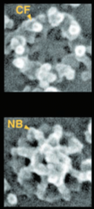 In the past 30 years the nuclear pore has been studied, as it is the gatekeeper between nucleus and cytoplasm. It seems to me that it is probably not the only way into the nucleus, and i freely say this from ignorance of nuclear pore biology, but just because in general, it is very unlike “mother nature” to provide only one way to do things.
Their figure Figure 2 from which I have cut and pasted a portion, is just beautiful, looking into the nuclear pore (using SEM) from the cytoplasm of the cell, seeing the nuclear pore basket below, and then looking up from the nucleus of the cell outward, seeing the basket in the center. What I was struck with is the relative emptiness of the areas between the 8 spokes of the nuclear pore, and immediately thought that the transport of small molecules could just go through there..but I would have to think that the pore would be unlikely to let past 10 nm molecules at the same time as a 39 nm molecule (i think that was documented in some study… especially since the total central pore unit is 45-50 nm. I can visualize this wacky wiggling of things being imported and exported through the center of the pore bouncing from side to side, letting smaller molecules go through when possible as larger molecules get transported through. Reminiscent of the activity in a pinball machine bouncing around.
A549 cell in apoptosis
A549 cell in apoptosis: RNAi knockdown of C9orf82 induced apoptosis in A-
549 cells. Huge granular center of the nucleolus with very little differentiation or dense fibrillar area is seen in middle right part of the micrograph (purple) and a part of the nucleolus with tiny light fibrillar centers and only a small amount of the dense fibrillar component. A portion of the nucleolus is off to the left near a nuclear pore. The cytoplasm of this cell has some RER that does have ribosomes, mostly near the nuclear membrane, and only three sort of not to clear mitochondria exist, one of which is apposed to an nuclear pore. There is very little condensed chromatin around the periphery of the nucleus, mostly euchromatin which is very coarse. Some perichronatin granules, and a little bit of interchromatin granule cluster (speckles) looking like a baseball cap above the circular structures of the nucleolus. Within, perhaps is something dense (don’t know whether to call this a paraspeckle or not (the location within the speckle fits this description). Nuclear chromatin and nucleolus purple, nucleus itself (which includes the interchromatin granule area) orange. 18272_78932_a549_4_si67_apoptosis
Civility
civility is a courtesy which is in short supply: Just a case in point here at the Rainbow room of CCHMC. Tall foreign obviously professional person (i suspect MD or research faculty) in a hurry to get food and go back to whatever he was doing, and maybe it was important, just kept doing the “following too closely” thing, as i paused and waited for other people to pass or backup from a counter. I moved over and said “go ahead, you are clearly pushing” he was pissed… but i just waited for him to go ahead…. he got soup… ha ha… which was followed by a rather vigorous thrusting of the ladle back into the soup pot which created a mess… i just said “oops” in a high tiny voice.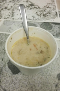
I wonder if he knew what a bad impression on all of the faculty and staff at children’s hosp that showed. Besides, it is a rude awakening for him… ha ha… he may know one small field of science very well, but he obviously does not know any physics, or he wouldn’t have tossed a large round bottom ladle into a pot of liquid…. oh well.
BTW, the soup (they called mushroom bisque) was terrible, too salty, and tasted and smelled like the inside of Pier 1 Imports. ha ha.
DEAR RAINBOW ROOM at CCHMC, that may be the nastiest soup I have ever eaten.
