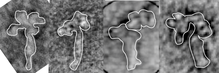
OK, apologetics first, i am not a protein chemist. But I ran this molecule in its entirety (it got truncated by the programs) so I diced it up into many many pieces, running each separately and trying to figure out how the pieces might fit together. This diagram (on the right) is the one I will use to create the octadecamer, the ball and stick diagram on the right is what i think is pretty accurate (and it matches some, but not all of the images I found in publications on SP-A, and it is just a little more accurate than those that make the CRD unit a round ball and the neck region a spring. It seems that only when I get the structure of the collagen-like portion along with the coiled coil of the neck to i get a structure that sort of fits what is seen with shadow cast TEM images (also published in peer-review journals). So this is what I am using. feel free to criticize. ha ha. 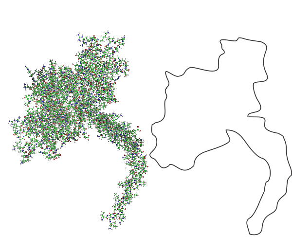
Here is a composite of several shadow cast images of SP-A, you can see that there is similarity. The dissimilarity with the ball and stick model might come from the fact that there are two configurations, open and closed, and that the closed configuration (which looks like the shadow cast molecules are in) is related to ion concentration. Almost all models of SP-A say there is an important “kink” in the collagen-like portion, and I am guessing that this is important for the images seen in traditional TEM of the alveolar type II cells which contain putative surfactant protein-A granules.
Monthly Archives: October 2017
Price reduction!!! ha ha still too much?
“In 1990, Congress established funding for the Human Genome Project and set a target completion date of 2005. Although estimates suggested that the project would cost a total of $3 billion over this period, the project ended up costing less than expected, about $2.7 billion in FY 1991 dollars.”
“But the price for genome sequencing has been in continuous free fall since the beginning. In 2006, Illumina’s first machine could sequence a human genome for $300,000, and in 2014 the company announced it could do the same thing for $1,000.”
“BGISEQ-500 Service Human Whole Genome Sequencing from $600, data delivered in standard industry formats”
If you are a —
If you’re free-thinking, they will think of you as a trouble-maker
but, if you’re a conformist, they will think of you as weak
——————————————
If you’re educated they will call you egotistical
but if you aren’t, they will call you stupid
——————————————
If you work but can’t support your family, they will call you lazy
but, if you work 2 jobs, they will call you a bad parent
——————————————
If you don’t have a car, they will think less of you
but, if you have a nice car they will think you’re pretentious
——————————————
If you stick by people that do you wrong, they will call you a fool
but if you cut people out of your life, they will call you disloyal
——————————————
If you are overweight they will call you unattractive
but if you diet and exercise, they will call you vain
——————————————
If you don’t do your makeup and your hair, they consider you ugly
but if you do, they consider you fake
——————————————
If you didn’t vote they will tell you were negligent
but, if you did, they will tell you it was for the wrong person
——————————————
If you were born with the help of affluent parents, you will be “entitled”
but, if you take help from others you will be a “freeloader”
——————————————
There are 10,000 ways to skin a cat.
In a world we have created, in which NOBODY is ever “doing it right,” wrong opinions abound, and everybody has one… but also, everybody has the right to be opini onated. WHO DOESN’T SEIZE THAT OPPORTUNITY!!
Just as long as that individual isn’t causing me personal problems, and mine isn’t doing the inverse… then let’s just ALL be the biggest that we can be!
Just do everybody a favor and CLENCH when you feel the urge to deliver MORE than JUST your opinions on people.
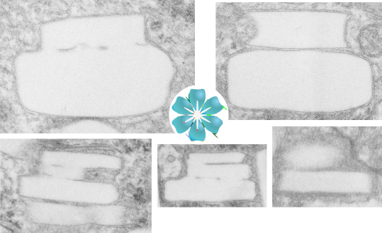
Wedding cake crystals: perfluorodecyl iodide
These crystals are certainly odd. I have for many years wondered at their layered cake appearance, and today just found it so funny that i had to make a spoof about it. So here are a few images taken from within a single hepatocyte phagocytic cell (like a resident macrophage) and these layered cakes ‘so to speak’ are oriented with the largest layer at the base and stacked vertically. The crystals appear to be stacked round layers, but probably not always, maybe with a chance orientation of smaller layers “up one”, I have not done any counting to see whether they might be “reducing in size as they stack”. The rounded bottom layer (not shown in these images) is seen in cross section sometimes as a full circle, other times as a tangential cut with one straighter edge and one rounded edge, just as one would find in a tangential cut of a layer cake.
At any rate, these are clues about the structure of perfluorodecyl iodide crystals in vivo.
Aspire to Inspire
Thoughts change reality
Listened to Bobby Schuller on youTube – saved these great words
 This moment of graphic sense brought to you by:
Spleen of a mouse receiving perfluorodecyl iodide emulsion–
The spleen, an immune organ, initiates immune reactions to blood-borne antigens and filtering out foreign material and spent circulating cells. It comprises at least 2 primary cellular compartments: the white pulp with its “periarteriolar lymphatic sheath, marginal zone and follicle” and the red pulp with its “reticular fibers, reticular cells, and associated macrophages”. These compartments have different vascular and cellular architecture.
Looking at mouse spleen after the infusion of a 10% IPFD 5% F68 emulsion at 100CC/kg, 55 days post, I had expected spleen to be completely packed with macrophages full of IPFD particles and yet I dont think (this is an estimate) that the volume density of crystals would be any more than 1-3% of the whole spleen. It is also clear that white pulp is pretty much devoid of cells with IPFD crystals, and IPFD crystals were pretty much limited to the red pulp. A quick look says that megakaryocytes don’t take up IPFD, red cell production, mitotic cells dont have IPFD inclusions (or much of it) that areas that are most likely to have cells with IPFD inclusions are around blood vessels. I have spleen tissue from infusion of many other prerfluorochemical emulsions…. spleens with IPFD are going to have to go to the back burner till I can figure out some fun stuff about liver.
It looks like IPFD does exit the liver after a period of 200 days or so, the crystals in the liver macrophages have largely disappeared, but what is left in the wake of the crystals is a host of cells that are in the interstitial and periportal spaces which should not really be there. The macrophages that were multinuclear remain that way with many dark areas (organelles) likely remnants of the massive lysosomal structures that were present during the IPFD occupation.
Kind of weird: mutate blend or die
You know…. i resent a little bit being moved from the building i worked in for 35 years…(that building went down and now there is part of a parking lot)(albeit a parking lot for those who should park the furthest away for their health but have close spaces to perpetuate their conditions)…. i survived the move; i resent a little bit being moved from the room I had for 6 years (ordered to leave with disrespect and uncaring spirit, though with a reasonable discussion would have gladly left)… i survived; i resent a little bit being given in an office where no one speaks my native tongue (english)…. haha (these are kids using the room where the emerita/emeritus faculty could still be productive)..ear plugs in, heads down on the desk, i am unable even to ask if it is OK to turn up the thermostat; no one understands what i say, no one says hello or good bye or good morning or please lock the door when you leave…. i have no “sound” enabled on my computer, never sneeze or cough or make noise…i’ve survived; Ha ha… how things at this university have changed. I am not going to comment on whether the change is for the better or worse. Change happens. This leads me to follow ….. my own advice from decades ago. MUTATE BLEND OR DIE
This moment of graphic sense brought to you by: 
Perfluorodecyl iodide crystals in vivo: big
Just reviewing some slides of liver of mice given infusions of an emulsion of perfluorodecyl iodide…. some of these crystals which completely fill to overflowing hepatocyte macrophages (multinucleated macrophages or macrophages fused into multinucleated giant cells following the foreign body response to IPFD infusion). Just saying here, some of the IPFD crystals are upwards of 30 microns long, and vary in width from 10nm to many microns. There is a very obvious directionality, always longer than wide. Iin terms of the actual molecular polymerization of IPFD, which is necessary to establish what is “wide” and what is “long” — that remains to be determined. But for now, these macrophages are about 90% “other” with half dozen nuclei in a localized cytoplasmic – crystal free zone together.
And regarding spleen, it has far fewer cells containing IPFD than I would have expected (just a relatively small number of reticuloendothelial cells show collections of crystals (compared to liver from the same animal which does have multinucleated cells full of crystals). So that is kind of interesting.
55 days after 100cc/kg of a 10% IPFD 5%F68 emulsion, the hepatocytes themselves are very well protected from inclusions of IPFD…. there are some probably, but not many. Hepatocytes themselves look nice, but the clusters of macrophages, without eliciting much of a fibrous tissue response are neatly circumscribed.
If…Then
I sometimes get the uneasy feeling that if i see a pattern in an electron micrograph that someone is going to say, ah thats just fixation artifact… and of course they are right. All testing is “fixation artifact” but its the purview of each observer to determine what is background noise and what is real structure.
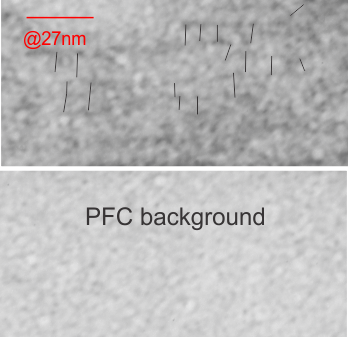
In the case of looking at perfluorochemicals left behind in tissues, consensus says that there is “nothing” in that empty footprint, just the embedding medium (in this case EPON 812). So there is a texture to the embedding medium, no question, but nothing I can perceive, and so easily compared to the texture of surrounding tissues, so that when I see a pattern in the tissue I am pretty convinced that it is the presence of the proteins . (They are influenced by the fixative and type and length of dehydration and other stuff not excluding the warping and shifting under the electron microscope. All those factors play a role). It is one of the reasons that an internal control for size (and i am suggesting background texture) in electron microscopy is really important. I CAN list a short list of variables to be aware of (my knowledge is just observational) which begin even before biopsy, including the time of the last meal, 1) circadian cycle, 2) health of the tissue (in vivo or in vitro –doesn’t matter) 3) mode of analgesia or anesthesia at biopsy (paralytics – solutions with ions (as in perfusion) 4) time till tissue gets into fixative, 5) size of the tissue block, 6) type of fixative, 7) length of time in fixative, 8) length of time and chemicals used for dehydration, 9)type of plastic, 10) section thickness, 11) staining compounds, 12) TEM specs, KV, etc…. so I am willing to accept that not all parameters are under one’s control.
I will however say that the pattern below (in a portion of a macrophage in the liver of a mouse given 10%IPFD in 5%F68, 267 days post infusion) shows a linearity I would not be willing to ignore. low mag micrograph top, for orientation, next below is micrograph of the red box area which shows the periodicity. That is just one of the linear arrangements that are visible in these tissues. 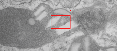
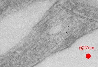
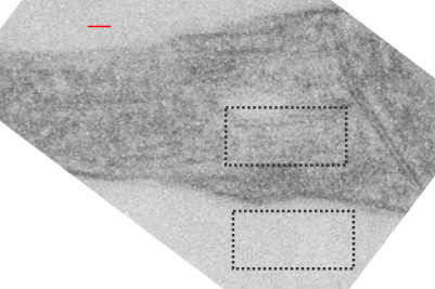 rotated image for easier measuring and comparing the textured are (top box) and the basic epon texture (bottom box).
