Category Archives: Electron microgaphs of lung

Various species of mammal and maybe a non mammal now and then have been obtained and examine using routine transmission electron microscopy. These are summary images.

Chocolate donut and sprinkles: nucleolar ultrastructure

Shapes from biology (especially microscopy) sometimes are reminiscent of the most mundane objects in our daily lives. This nucleolus has reminded me of a donut with sprinkles for several decades.  In searching the nucleolus for clues to the connection between the sizes of apparent granule-fiber, beads on a string, things to what has been reported in the molecular structures online, I used this particular nucleolus (from a baboon that had been given perfluorophenanthrene emulsion (probably IV)) and some tissues biopsied. This is lung. In other views, this particular cell (not clearly identifiable but likely an alveolar macrophage) contains PFC droplets.

The “sprinkles” for this donut represent the dense fibrillar component (entities measured on a post from yesterday; the cake part of the donut is represented by the circular structure in the center of the side-by-side identical micrographs, which is the granular component. NB, the granular component on the left is light grey and round with the fibrillar center being the center of the donut.  It was noted in some of these nucleolar structures that there is such definite banding and that the lumps on a string is a very good casual description of the organization of RNP along a template and the measurements are pretty good.  As a reference point the diameter of a ribosome is about27 nm and the space between ribosomes in this particular cell is around 58 (or 2x the diameter of a ribosome). The diameter of the beads in the granular portion is 21nm, with a distance separating them of from adjacent structures is quite large and prominent, around 42nm (twice the diameter of the granule and though smaller in overall size than the ribosome and its space) is quite similar to it. The granules on a string in the dense fibrillar portion of the nucleolus is close to 30nm, slightly bigger than the average ribosome, and the space around these larger granules is the smallest among the three, at 38nm.

A second organization to the granular portion is seen in the parallel alignment and even spaceing of the granue. Orange lines lie over the very obvious linear pattern in the granular center of the nucleolus. A similar linear alignment is found in the dense fibrillar portion which becomes very very obvious in cajal bodies (not seen in this micrograph).
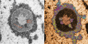
electron micrograph of the nucleolus of a baboon lung cellThis image would be a fun cover submission…  maybe someday i will make up a panel with three or four TEM look alikes. I know i have a valentine, and a ghost and a donut.
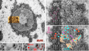
Granule sizes and spaces in the nucleolus

Epithelial cell in the lung of a baboon (probably a type II alveolar cell) showed a ring nucleolus with a single fibrillar center and concentric granular and dense fibrillar components. I took the time to measure (relative to a series of ribosomes measured in the adjacent cytoplasm – estimated at 27nm in diameter) the size of the granules in the dense fibrillar portion and in the granular portion of this nucleolus.  The image on the left shows the whole nucleolus, the three divisions (there are probably more) of the nucleolus are prominent. From that image of the nucleolus, the orange area is enlarged twice on the right to highlight distance between granules of the dense fibrillar component and the granular component. The small redish box is what was used to estimate the size of those granules by measuring ribosomes therin (red dot) obtained from a portion of adjacent cytoplasm from the same image.
The diagonal (and rearranged and measured as horizontal) lines are those between granules in the granular portion (white) and also the dense fibrillar portion (purple).  The spaces between the granules in the granular portion are marked out with a peach transparency and the blue circles represent the size of granules in that area.

Key: orange circles = size of the granules in the dense fibrillar portion at 30nm, with about 38nm distance between them; white lines and blue circles  21nm) represent the diameter of the granules in the granular portion and the distance between them (42nm).  For comparison, the ribosome (red dot, lower center-left is 27 nm, and the distance between ribosomes is about 58nm. The apparently-coiled strings (pink) are something that occurred regularly, kind of an arcing gently oval beaded appearance. 13279_94-32_baboon_lung_PFP (perfluorophenanthrene).

electron micrograph nucleolus monkey lung perfluorocarbon

Pseudo-colored alveolar type II cell granules, and unretouched, and composite

Pseudo-colored alveolar type II cell granules, and unretouched, and composite image.  I am just about finished with this study. I submitted the manuscript, I do hope if flies without too much revision. I am very aware that it is totally descriptive, but then descriptive science was the first science and it has great value. We all learn by what we observe, more so than what we propose (hypothesize) and test.

Here is a granule from my fav guinea pig #301 neg 9837, block 17084, which has several stacked parallel periods of the layered granule all in pretty nice array perpendicular to the layering. I extracted and then rotated the cropped portions, and increased contrast, and added half transparency to these four seemingly ideal areas. I merged them (with their transparencies matched as best I could) and flattened them as a fifth inset.  This image is the inset in pink with 100 nm markers vertical and horizontal.  The individual crops are shown below, colored just for fun.

layered protein in alveolar type II cells

electron micrographs of alveolar type II cells in lung guinea pig

..yes, one more cover submission illustration — I just love these granules

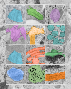
..yes, one more cover submission illustration — I just love these granules, and I think this might be my favorite yet. Colored all the granules in (using photoshop) overlying their own original images from the basic research on this putative SP-A granule, and arranged them in a highly ordered format (haha) which is certainly not really my normal MO, since I am kind of a random thinker.  But this is nice…in my opinion, and also a little artsie-craftsie for a scientist.. but thats what covers for scientific journals are for.

…and yet another cover submission for the manuscript on SP-A granule and electron microscopy

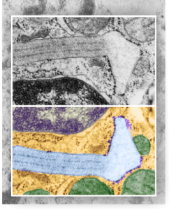
…and yet another cover submission for the manuscript on SP-A granule and electron microscopy.  This one is a little more dramatic than yesterdays post from the bottom right part of the image.  I like it better. It shows the quintessential granule, in black and white at the top, color at the bottom in a vertically mirrored image, directional ribosome attachment, electron-lucent areas under the non-oligomerized protein part, distinct periodicity to the dense bands (three complete “periods” in the lengthwise portion of this granule) and also the periodicity to the dense layers (3-4 dots/100nm) and even some of the faint periodicity found in the central lighter layers. In this electron micrograph one also sees part of the nucleus (bottom), a couple of nuclear pores, and some perinuclear space (but no granule formation in the perinuclear space is seen in this image, but it is found there frequently) and on the top, portions of three mitochondria and one adjacent to the lucent area of the granule. There are also other profiles of RER, coming and going which at some point might connect up to layered putative SP-A protein granule structures.

Four cover submissions for the SP-A manuscript: love the colored electron micrographs of granules

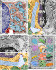
Four cover submissions for the SP-A manuscript: love the colored electron micrographs of granules. I am submitting this to Anatomical Record. They request a cover submission at the same time as initial manuscript submission, apparently, at least those are the instructions.  Typically I think the manuscript has to be accepted before cover submissions are made.  Oh well, I am just hoping for the best.  Preview here.

3 diagrams as putative fits for alveolar type II cell granule

So tired of looking at this structure and trying to brainstorm about how things fit together and whether SP-A is a good protein fit for this very regular and very nice molecular alignment within the RER of alveolar type II cells of several species.

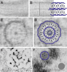
A. Single period (RER membrane on top and bottom) from ferret show outer dense layers and less dense central layer. B is also a single period of a granule from guinea pig which is not bounded by RER but by another period above and below. Less electron-density accompanies of outer dense layers occurs when adjacent periods are present. C is an end on view of a cylindrical granule from guinea pig, almost 200 nm in diameter which could be twice the single period width. The inner dense central layer would be compressed into the central density. The RER membranes are seen on the upper and left on the outer edges, and periodicities for both the less dense center layers and dense layers appear as concentric rings. D, is the same image as C, with a cylindrical model of an end-on view of the possible arrangement of SP-A molecules which could account for this structure. The number of densities in the concentric layers was used to calculate how many molecules to use for the model. Vertical lines (shown in Fig. 5 as well) became radial spokes. Bars = 100 nm. E. On rare occasions, and only in guinea pig, round and dense fuzzy-ball like structures were seen squeezed between the typical 100 nm periods. Box in E was enlarged in F to show the relative sizes of the round densities. A diagram of a potential SP-A fuzzy ball, approximately 30-40 nm in diameter approximates measurements of the spherical densities. Grey circle is ribosome size for comparison. Bars = 100 nm

Diagram: SP-A and lamellar bodies

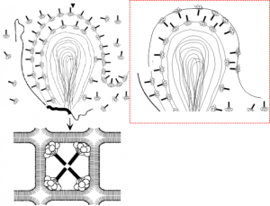
An article by Palaniyar N, Ikegami M, Korfhagen T, Whitsett J, McCormack FX. 2001  entitled Domains of surfactant protein A that affect protein oligomerization, lipid structure and surface tension, published in Comparative Biochemistry and Physiology 129:109-127, presents a diagram of how the 18-mer of SP-A might integrate itself into the outer lamellae of lamellar bodies in alveolar type II cells.  I did notice an inconsistency, perhaps, in the diagrams. The bottom diagram (left bottom) shows the SP-A molecules with their N terminals all pointing inside, or towards each other in the center of these tubular myelin structures (the diagram is a 2D model of a 3D structure, hence “tubular”).  In the granule found in alveolar type II cells which I have been trying to describe morphologically for several years has a layering pattern that suggests that the carbohydrate recognition domains are actually pointing “away” from each other.  This is replicated in the diagram to the left.  The inconsistency comes in how the SP-A molecules are represented in the top portion of the diagrams, which I duplicated in part, and re-oriented the SP-A molecules to match the tubular myelin arrangement shown in their figure (bottom left).  To me this makes more sense, and it also is consistent with the layering of the alveolar type II cell granule in guinea pit, ferret, and dog, and in addition, with the outward orientation proposed for the Birbeck granule (the C-type lectin, langerin).  Comments are welcome. My edits to their diagram are in the red dotted inset to right.


SP-A: three possible configurations to match electron microscopy of alveolar type II cell granules

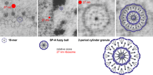
Still working on this granule in alveolar type II cells. I think there are three levels of organization to the cylindrical and circumferential manifestations of whatever surfactant protein comprises these granules. The original 18-mer, well confirmed by numerous other scientists, on the left, relative to the size of a ribosome, and also with the hexagonal order seen in tangential sections of the outer dense layer of the granule. Middle image is the proposed SP-A fuzzy ball which seems to be a spherical organization of the same 18-mer, just with something different happening in the center region of the sphere. I can’t even guess what that organization (missing perhaps some of the CRD regions of SP-A?) to make things fit. This image also relative to the size of a single ribosome (red dot). Then on the right hand side is a double-layered cylindrical granule (this one likely from guinea pig) which has a diameter of about 200 nm, which means is likely has a column of four molecules for each radius. Size of the actual superimposed cylindrical period, and then a relatively sized – diagram…  sized relative to the other spherical or concentric configurations of SP-A.   The micrographs do pretty much dictate the arrangements, or at least that was what dictated the arrangements of the SP-A stick figure I used (which was vectorized from models of SP-A in publications.

A better fit model for langerin and profiles of Birbeck granules

Lots of images have been published for Burbeck granules. I am interested in them only because they seem to be the only other C-type lectin which has a substructure that is organized and obvious when viewed with electron microscopy. Well not the only, because I think that SP-D has fuzzy ball structure that is seen with TEM. It is puzzling why MBP and some other lectins don’t assemble into orderly granules like langerin, and SP-A (at least I think it is SP-A).

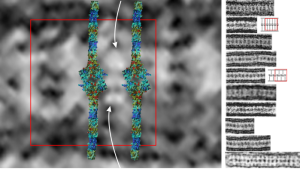
So here is a composite that I have made from TEMs posted everywhere in the literature, lined up with the 50 nm distance between the ER membranes, and such a very predictable substructure of proteins organized within.  One publication in Plos ONE (Glycosaminoglycans Are Interactants of Langerin: Comparison with gp120 Highlights an Unexpected Calcium-Independent Binding Mode. Eric Chabrol, Alessandra Nurisso, Antoine Daina, Emilie Vassal-Stermann, Michel Thepaut, Eric Girard, Romain R. Vivès, Franck Fieschi) gives a molecular structure and a hypothetical overlay onto TEM images.  I think they missed it by a little…. for starters their intramembrane part of the molecule is NOT shown in their diagrams, which makes the molecular fit difficult since the CRD of the molecule are a relatively larger portion of the actual TEMs than is seen in their molecular diagram (line diagrams on the far right of the image). Additionally, there is a cytoplasmic portion of langerin, at opposite ends of the mirrored CRD portions of langerin which their diagram does not show (don’t know why they chose not to show it).I vectorized their diagram and have placed it back in the 50 nm membrane boundaries of the ER with the transmembrane portions actually existing and that makes the size ration of the model molecule fit the actual TEM much better.

Again, my only interest in this is that I am trying to ascertain whether my thought that SP-A is responsible for the granule in guinea pig and ferret type II alveolar cells true.

Right side of the diagram is a collection of Birbeck granule TEMs sized to 50 nm, the image on the right shows a red 50 nm bounding box, and four langerin molecules oriented CRD inward, and transmembrane areas through the ER membrane.  Just my opinion here but there is another molecule or some portion of langerin which is “between” the four diagrams superimposed. White arrows point there.  Their diagrams (two alternatives) are shown in red and grey to the right of the actual TEMs of Birbeck granules.