Monthly Archives: August 2016

Smooth ER between RER lamellae in guinea pig alveolar type II cells

This electron micrograph shows three very long (a portion pictured here) lamellae of RER with a surfactant ?? protein, maybe surfactant protein A accumulation which shows a very very light area of banding – ICB) , and sandwiched inbetween these three are flattened cisternae (black arrows) which do not apparently contain the protein accumulations that is present above and below each.  Whether these flattened cisternae without apparent protein are going to be sectioned down the block as thickened lamellae with protein content, is up for grabs, but likely (see lowest black arrow which points to a thickened part of the flat cisternae).  This configuration is a little unusual.  Other things to observe in this electron micrograph are: part of a lamellar body – upper right;  tiny hint of the central band typical of what I think is overproduction of surfactant protein A – red arrow; basement membrane beneath the alveolar type II cell, and what is probably an endothelial cell – blue arrow;

icb_10254_gpig

Just 4.5 million posts on “spatial reasoning”

So here we have a subject of great importance and reasoning.  First quote from a google search says “Spatial reasoning is a category of reasoning skills that refers to the capacity to think about objects in three dimensions and to draw conclusions about those objects from limited information. Someone with good spatial abilities might also be good at thinking about how an object will look when rotated.

I can attest first hand that some individuals are totally gifted in this area per the following:

I requested from a local lumber milling company 100 blocks of different species of wood, planed at 1/2 inch and cut in increments of one inch in all permutations and combinations. Such as 1×1, 1×2, 1×3, 1×4, 1×5, 2×2, 2×3 2×4 etc etc…  The mill-man told me that something under 8 inches was possible with standard wood. I requested as many cool scraps with knots and stains and odd grains and mineral deposits as possible, just because those are visually interesting to me.  When I picked up my order, I was thrilled, but also totally overcome in awe of whomever had packed that box of pieces. Well over 300, perfectly placed in a box, few spaces, all assembled just as one would assemble a jig-saw puzzle. I do not have any idea how in such a short time anyone could have created layer after layer of even fitting wood using random size pieces in such a short amount of time.  Astounding spacial perception is all I could use to explain it.

Spatial reasoning is usually classified differently than other forms of reason: verbal, logical, and associative skills and working memory.

Spatial reasoning abilities allow individuals to see, remember, rotate, mirror, invert objects in their minds. While in times gone by, men were considered better at spacial awareness, this and other discrepancies between the genders has been challenged. Sadly, as with most literature, there is suggestion that age takes its toll on spatial (as well as other) reasoning abilities. But on the upbeat side of this, many views of the “non modifiable brain” have been disproved, and the mind and cognition are recognized as powerful, plastic (you know not in terms of PVC plastic, or polymer clay,  ha ha) but malleable, which gives us hope and maybe impetus to improve our mental abilities.

It isn’t a matter now of “right brain” “left brain” but networks  (sound familiar to you computer scientists).  Networks that take care of “executive functions”, that take care of “creative and associative” problems, and what i read was called the “default mode network” (ha ha stay out of that one–involved in inhibitory control), and the “salience network” making decisions about whether to stop, redirect, emphasize or continue and partly in control of the default mode network… at least that is how i understand it.

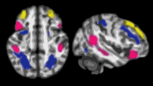
So when I looked at the color MRI images of brains functioning in these different modes, it became clear that there were “hot” spots placed at several different sites in the hemispheres, distributed as it were, and while probably not redundant, certainly intertwined, interlinked, and interacting.  Not just one hemisphere and one active spot, but many, linked.

The studies that show up on function of these networks seem largely to focus on tranmatic brain injury. I did find one study that showed (MRI) the distribution of some areas of activity for several networks.

I put together a composite color image (using photoshop and corelDraw) from several MRI images that I found online…. I think this is an awesome and fun graphic…. we are wired–right and left hemispheres. I guess, most importantly for society as a whole, the frontal lobe networking (the salience, the breaks, the control) (yellow) (my image modified from a Scientific American article, which by now is probably highly outdated… LOL)

brain_networks

There is no “get out of consequences free” card

So here is a thought.  Why does one do things that are bad for one’s own health for 45 years, then expect to change suddenly and have their health improve overnight.

get_out_of_consequences_free_cardIf one has abused one’s own physical body with smoking, or drinking, or sloth, or eating too much, but at age 66 suddenly change lifestyle behavior (quit, abstain, tidy up, eat right, you might think that it is appropriate for the healing to take place in a matter of a few weeks.  Biology isn’t like that. If over a 50 year span one has been neglectful, what gives us them the right to expect healing overnight. Forgiveness is a moral issue, there is no forgiveness in biology…. but there is repair, so repair of cells and tissues is biology takes time, a long time.  There is no “get out of consequences free” card in biology.

  1.  Doctors (even though they tend to think this) cannot save everyone. Nor should doctors go about the business of pressuring us into having procedures which are good for the “bottom line in profit” but ultimately may affect our health negatively. The same is true for Big Pharma, telling us that we should medicate every ache and pain (largely stemming from our inactivity and innatention).
  2. We make choices (the up quark and down quark (physicists, forgive me) minute by minute, hour by hour, and so on, and even attempting to do what is biologically right may not work (because of overriding and as yet unclear processes).
  3. We can repair, this is a good thing, but repair doesn’t come to those who hide.

Fuzzy protein granule in rough endoplasmic reticulum of a type II cell

Original, and unretouched or processed (not even contrast enhanced) photo of a small profile of rough endoplasmic reticulum from the alveolar type II cell. It has two ribosomes over to the left about 10 o’clock, they can give you a feeling for the dimensions of the fuzzy balls within the central portion of the profile.  The electron lucent area surrounding the dense center is pretty common in these intracisternal bodies (granules if you prefer) and the layering here is too large to be part of the 100 nm pattern seen when the bodies are found perpendicular to the periodicity.  I looked closely at the center, and I would estimate that the bodies (in groups of three – yes, my guess – roughly) is something around 60-70 nm, at least bigger than the ribosomes (when lumped together in threes or whatever the cluster contains.  OF course the whole granule is much bigger, but it would be fun if the smaller portions of the central density of this intracisternal body equated to 50 – 60 nm groups of three bouquets of surfactant protein A.  That would be just fun.

fuzzy_balls_single_all_grey

You can find a short animation with text at this link to YouTUBE:

Change: resistance to change

Sometimes we meet change (at work, home, the fruit market, anywhere) with resistance.  I googled to see if I resisted change more because I was “old” (as is the case).  I was amazed to find out from an article written by people in Switzerland (albeit just one paper) that older adults are actually less resistant to change in the workplace.  So that was really fun trivia to know.about_change

I also am happy to say that I am not the only blogger in the world who makes typos, grammatical errors and doesn’t find the time to correct them either. So here is a summary statement of what I find about change, and noted that the bulk of what google fed me related to managing change in the workplace, for the most part which addressed “before – change” strategies helped the most at maintaining calm.

For me, change requires much effort to overcome.

silence does not equal consent
change happens
change is not always good
change brings chaos
change brings order
no change = no growth


More periodicity seen in RER granules in alveolar type II cells

More periodicity seen in RER granules in alveolar type II cells from a ferret. In the areas of this single intracisternal RER protein there are great examples of the periodicity of the protein as it was fixed (this fixation was standard paraformaldehyde glutaraldehyde fluid). This particular electron micrograph is a little grainy (I probably used dektol in stead of microdol to develope it — in hindsight not a good shortcut to take, but sometimes we do things without knowing the 30 year consequences.. duh.  At any rate, I have compared areas in the dense band with the lucent bands of this particular tangential cut through a curved RER granule also called intracisternal body and shown the unretouched (in terms of the densities) and the burned densities as I could see them.  There is a portion where there is not periodicity taken from the exact same RER granule less than 100 nm below the string of beads seen above it.  Ribosome (20-30 nm? is used to compare sizes, and the beads look to be something on the order of 50 nm.

Full granule is pictured in the transmission electron micrograph on top, arrows point to the horizontal segment which has the unretouched periodicity, the arrow below points to the lucent zone used to compare whether I am hallucinating the periodicity (ha ha).  Micrograph 2, just beneath the full granules is enlarged unretouched (i did remove some dust and a scratch using the stamp tool in photoshop but in the line of granules). Micrograph 3, is a view of the exact same strip of the intracisternal body as image 2, but i enhanced the beads with the burn tool. and micrograph 4 is the more or less no-granule-zone in the lucent area as spread out in a tangential cut, unretouched.

more_p[eriodicity_RER_proteins