Monthly Archives: September 2016

Alveolar macrophage: 3 hours liquid breathing E2, 17 days recovery

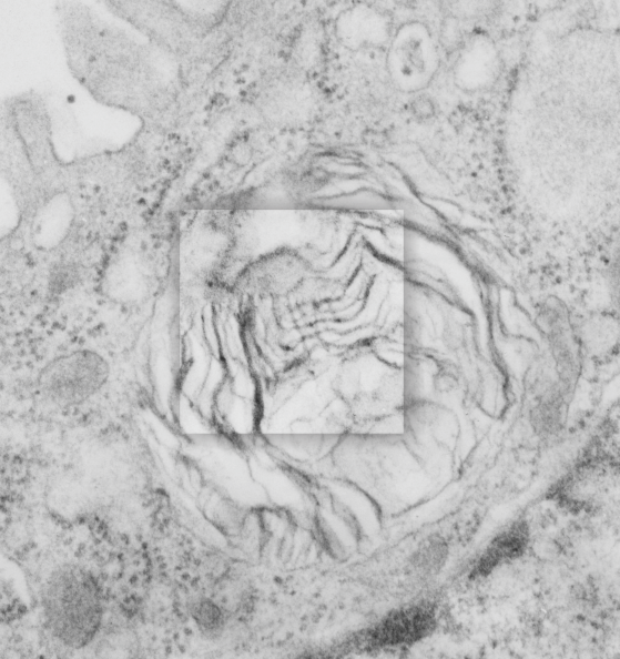
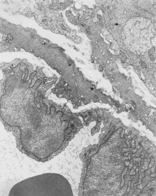
Alveolar macrophage from a mouse which was submerged in perfluorochemical (PFC) liquid and which breathed that oxygenated fluid for three hours.  The mouse was allowed to recover for 17 days, then the lung was preserved for various chemical assays and for transmission electron microscopy. This experiment took place on 2-1-1975 in the laboratory of Dr. Leland Clark, Jr, who was investigating perfluorochemical liquids as possible remedies for respiratory distress and for alleviating bends in deep sea divers.  This original micrograph, 10,000 x at the microscope, enlarged 4 times is my neg. 1436, block 4820.  The original micrograph can be scanned and sent to any one who desires it.  The most obvious anatomical markers are the nucleus, which looks a little like a pac-man mouth (LOL) and it has an active nucleolus, a few not so opportunely sectioned nuclear pores, some golgi vesicles in the cleft, occasional profiles of RER, about a dozen mitochondria are present as well.  There are two structures which are NOT likely to be inclusions of E2, since they have other lamellar and whispy electron dense contents.  Those circular droplets which I believe represent the footprints of E2 are colored (left) and left original color (right) in a contrast enhanced but otherwise unaltered micrograph.

1436_4820_E2_lung_liquid_breathing_macrophage_TEM

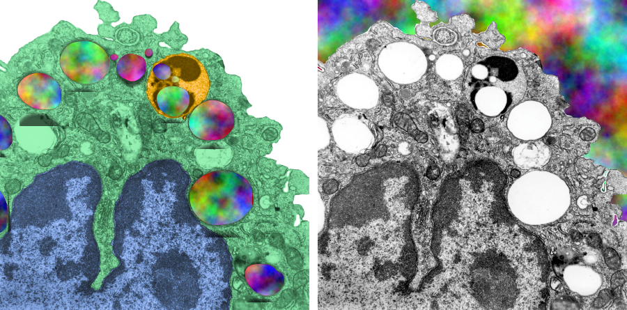
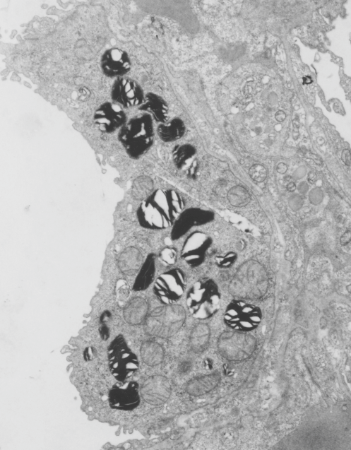
There is a dense body (actually 2 dense bodies) which have E2 inclusions, the presence of PFC droplets within membrane bound lysosomal bodies accompanied by dense enzymes was common for just about every pfc that was examined, though the formation of those lysosomes (dense bodies) took several days to weeks to form.  There is really nothing too remarkable about this alveolar macrophage (as far as I can tell) except for the presence of PFC. Of course I had fun with colors here, nucleus, blue; one dense body/lysosome, orange; alveolar macrophage cytoplasm, green, in left image.  Right image, contrast enhanced original.

Lamellar body in an alveolar type II cell has surfactant-like-tubular-myelin grid pattern

Lamellar body in an alveolar type II cell has surfactant-like-tubular-myelin grid pattern. This was found in an electron micrograph of a ferret type II cell (neg 9872, block 23462, ferret # 7, control, 6 10 1982). This particular lamellar body is unusual in that within the lamellar structure, there is a grid like pattern which is normally seen only in the alveolar space. While, according to the literature, alveolar type II cells can internalize mature surfactant from the alveolar space, this doesnt anatomically seem very likely to me as an explanation for this particular structure.  I thought it was unusual enough to post. I put a bounding box and a drop shadow around the area of interest.  The grid inside has at least 4 partitions horizontally as well as vertically.  Surfactant protein A is likely responsible for the grid (according to consensus) though this is usually in the alveolar space. This mineature tubular myelin was described in a book, ed Jacques Bourbon I think, which was reported to be about 1/3 the size of tubular myelin in the alveolar space.

9872_23462_ferret_lamellar_body_grid

Here are two reference images for the above micrograph to give approximate sizes. Regular tubular myelin has a side to side distance of something around mini_tutular_myelin_in_lamellar_body1100 nm, whereas this intra-lamellar-body mineature tubular myelin sample is about 60 nm?  Ribosomes are 25-30 nm (black circles)


Alveolar type II cell RER inclusions: two different proteins? two different organizations

THis particular alveolar type II cell (from one of my most favorite guinea pig blocks, i.e. # 301, comes this profile in which there are two enormous RER bodies (at this point I don’t think it is fair to call them granules, but something else, I don’t know what) which are upwards of 20-30 microns across.  One (on the right – see inset box) has the organization of a usual intracisternal body or granule that I have repeatedly found in guinea pig, at least the animals that were in our experimental group back in the 1980s) and the other does not.  I am not bold enough to call this a separate surfactant protein type of granule (as opposed to what i think the right hand granule is, that is, surfactant protein A) but it certainly has its own unique banding.

9817_17084_gpig_301_two_ICB

Electron micrograph after liquid breathing PP5 for 1 hr, 1/2 hour recovery

Mouse lung, here is one of a few of the electron micrographs taken from lung tissue after various numbers of hours of liquid breathing,  in this case the fluorocarbon is PP5, the duration of time was1 hr,  with a 1/2 hour recovery. The negative is my number 1489, block number 5473, just in case there is interest in this image I can provide a higher resolution image or some additional backup.  I recall that some of the perfluorochemicals used in the liquid breathing experiments seemed to cause a clumping of platelets in the very small vessels in the peripheral lung.  I was struck by this early on, and I did not occur with all PFC, though I will need to confirm this.  This particular mouse (4-7-1975) lung showed inclusions of PFC, and displayed some of the vascular characteristics that I have seen with liquid breathing experiments before: specifically, the accumulation of platelet aggregates in the small vessel lumena.

This cropped area from a micrograph just focuses however on the two alveolar type II cells which show two anatomical characteristis which I think might be related to liquid breathing of highly oxygenated fluorocarbons, 1) the lamellar bodies are very compact (maybe even categorized as slightly “immature” 2) the mitochondria look like they are on “speed”  ha ha.  wide mitochondrial matrix areas and cristae pressed to the sides and possibly reduced in surface area.  Those two ideas are reserved for a later date.

This micrograph is a little out of focus, and is highly magnified from the original, quality is admittedly not great, but it demonstrates the point.  The initial purpose was to look for the incidence and morphology of intracisternal RER granues in alveolar type II cells of mouse: (none found in these randomly saved micrographs).

1489_5473_mouse_lung_PP5_1hr


Electron micrograph of mouse lung after liquid breathing E2 for 3 hours

Feb 19 1975 under the auspices of the Children’s Hospital Mental Retardation Research Foundation (no longer in existence) house in what was called the IDR building (Institute for Developmental Research – also no longer in existence) with Leland C. Clark, Jr, I was in charge of looking at histological changes from his experimental procedures using perfluorocarbons: as 1) possible components of artificial (or synthetic) blood, 2) for contrast agents in lung, 3) and for lung lavage and aeration fluids.  From the collection of animals and micrographs, here is a portion of a red blood cell, taken from an awake swiss albino mouse who had breathed E2 for 3 hours, and allowed to recover (apparently uneventful) for 1/2 hour.

I am totally not sure if anyone in the world will care about these data, but could not in good conscience allow some of the images to go into the recycling bin without recording them for posterity.  Given long life, I will at some point assemble them into a manuscript, to submit “where”  ha ha. Just relating this to the presumptive SP-A cisternal bodies (granules) that I am working on, none seen in this particular mouse (11 micrographs with type II cells at sufficient magnification to assess the RER and the perinuclear membranes.

Neg 1401, Block 4960, mouse 18.63 g, mag at scope=8,000 x, enlarg=2.83x, Karnovsky’s fixative instilled in trachea, osmium post fix, EPON 812 embedment.  Original description of section with LM = general increase in cytoplasmic lucency, no plately plugs, blood vessels (per this photograph) were mostly open.  Not seen in this micrograph but present in others to come, surfactant hypophase was abundant, the cytoplasm to nucleus ratio seemed increased, no E2 inclusions were found in alveolar macrophages.  Red cell within blood vessel at bottom, endothelial nuclei diagonally across middle of micrograph. Plenty of “transforming fibrils” as Dr. Lowe and Dr. Charles Basom used to call them are seen in the interstitium.  No alveolar type II cells in this image.

1401_4960_mouse_lumg_ E2_pfc_breath_3hr


Surfactant A protein and bilayers: does this in vitro data match in vivo data?

This research study uses what they call surfactant A filamentous structure and lipid bilayers to study whether surfactant protein A is responsible for some of the functional and structural features of surfactant.  In one of their figures I tried to match their banding pattern with that found in vivo in the various animals I have looked at searching for an identity for this RER granule (or intracisternal body) and the numbers don’t match up completely.  Their banding in vitro between surfactant protein A filaments and lipid bilayer material is more than the 100 nm periodicity I find in vivo in guinea pig and ferret and dog alveolar type II cells.  I am certainly not challenging their measurements, I would more likely challenge my own, since I have not photographed a grid pattern in decades with which to calibrate the TEM.  SO, that said, I conclude that there is the possibility that something in surfactant A is at work here, and the observation which is much more interesting is that they describe intersections at right angles and branpalaniyar_surfactant_A_in_vitroching in vitro… just like the branching and curving that is found in the granules (intracisternal bodies) in vivo.

Nades Palaniyar, Ross A. Ridsdale, Stephen A. Hearn, Yew Meng Heng, F. Peter Ottensmeyer, Fred Possmayer, George Harauz American Journal of Physiology – Lung Cellular and Molecular Physiology Published 1 April 1999 Vol. 276 no. 4, L631-L641

This is a crop and edit of one of their pix.  The white 100 nm bar is mine, made from their scale marker of 50 nm.


Cover submission to JBC: July 22 2011 made for Aziz Sancar

Little did I know when I made this for a friend of a friend that it would be for a Nobel Laureate.  This image is about biological clocks.  I did several different styles, but this one was accepted and published July 22 2011: Vol. 286, Num. 2.  I have no clue what made me swirl the clock faces into the columns.  Ha Ha.  But I am glad someone thought it was a good visual representation.  If I am not mistaken, this was a freebie, and in addition JBC didn’t put my name in for credits…. so much for fame and glory in the world of scientific illustrating.

jbc_cover_2011_SancarSo here is one submission for the same journal cover place as above, which was obviously rejected.  I agree with their choice.

jbc_cover_2011_Sancar_not_used

And… here is yet another cover submitted to JBC in competition for the same article.

jbc_cover_2011_Sancar_not_used_2

Cover submission @2010 to Oncogene: Apoptosis, snowflake

A panel of apoptotic cells in culture, fixed for electron microscopy, sectioned and stained, photographed and then played with in photoshop with a vector image added (created in CorelDRAW) with a light fill in the hexagon with different hue saturation and lightness from the original.  Wish of course that this had been a cover, but as is my experience, artsie and sciency only mix in the most elite circles.  Ha Ha. As I recall, i remade this for Microscopy Today without the snowflake, slightly less interesting, but a cover nonetheless.

snowflakes_apoptosis_oncogene_submission

Microscopy Today: Cover for January 2008, apoptoses as snowflakes

This was a cover that I had hoped would be published with an article that I helped to co-author that described a pro-survival gene, or otherwise functioning as an anti-apoptotic gene.  Playing with the images of apoptotic cells in culture I did manage to submit the image to Microscopy Today, and they published it in 2008.

I thnk what amazes me is that I really made a whole lot more cover submissions than were successfully published (LOL). I find a similar statistic in publishing data, where really only a small part of what is studied, learned, documented, and put out there in the world of science, makes it into print.  Good things have to get strained through the teeth of overworked and sometimes not that interested peer-reviewers.  Who, I wonder, thinks so highly of themselves that they have the right, and/or mission, to exclude the works of others.  Notwithstanding the well know fraud in science, the great majority of individuals working to find “truth” in all its glory, are honest and well-intentioned.

snowflakes_apoptosis_Microscopy_Today_jan_2008_