Monthly Archives: October 2016

Text book pictures of untreated and ozone exposed guinea pig alveolar type II cells

There is a book called Diagnostic Ultrastructural Pathology – A text atlas of case studies. Volume 1 ( Ann M. Dvorak, Rita A. Monahan-Earley) which on page 218 posts figure 182 (reproduced here – I thank them). It shows clearly that, while they were interested in lamellar body ultrastructure before and after ozone exposure, you can be sure that the most important structure for me is the “cisternal body” that flattened and layered protein structure that I see in the upper right-ish corner of their micrograph.  Guinea pig must have some very robust mechamism for responding to environment, producing proteins (presumably some surfactant proteins) and in this case, after ozone they do not seem to become more prominent (according to this chapter in the book) as they do in dog (see previous post), but actually become less conspicuous.

Dimensions and position within the cell are classic “cisternal body” which I really wish I could rename SP-granule.  Red arrow points to the RER granule.

diagnostic_ultrastruct_lung_book_guinea_pig

Eureka, “ah ha” !! I found reference to a study on dog lung which shows alveolar type II cell granules (cisternal bodies).

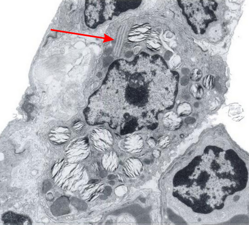
There was a group of scientists in california (I could not remember which research institute) that kindly sent me some plastic-embedded tissues of dog lung, since I had found a paper someone there that described layered protein granules in alveolar type II cells like I had seen in ferret and guinea pig and had ask them for some tissue. I could not (after 35 years, remember their names).  HERE IT IS!  awesome, though at least one of the researchers is deceased (J. Stara).  I am taking some of their electron micrographs, cropping, editing contrast (only) and placing them next to the ones I found in guinea pig and ferret.  Perfect matches.  This publication is so old (1973) that there is no way that surfactant proteins could have been implicated as possible contributing factors to these granules that appeared in their ozone treated beagle dogs.  The surfactant proteins had not yet been described.  Ozone caused what was described (for lack of background information on overall type II cell function in those days) “metabolic alteration ….. this toxic agent”, which of course is a statement which NOW in 2016 in light of decades of research on surfactant and lung function looks patronizing.

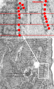
Their RER measurements made this “approximately 754 A (75 nm) is not that far off from what was calculated by my own measurements 100nm (microscopes differ in magnification tables, and so neither is likely to be exact).

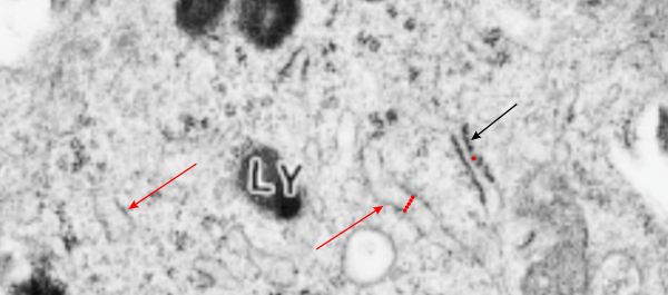
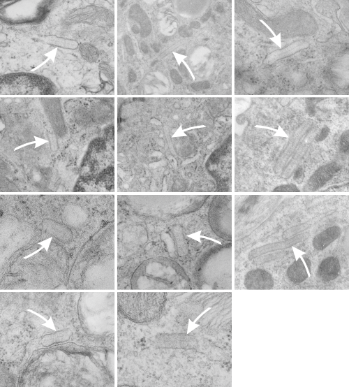
Because of their reporting of small, sometimes stacked “bar like” structures such as I have seen in untreated dogs (and looking a lot like what I have seen in rabbit type II cells).  it is likely that an “excessive” amount of surfactant protein is produced and maybe stored in a multi-meric configuration) within the RER awaiting utilization in assembly within lamellar bodies or multivesicular structures. Stevens et al, 1973, mention (yippie) that there is a dense line which runs parallel to the length of the RER in these infrequently seen structures.

Ribosomes in the beagle exposed to ozone that border the RER profiles on the growing ends of the layered protein do look to be in similar positions with regard to banding of the granule to those found in ferret. My inclination is to think that there is a ribosomes on either side of the central band in each period (beagle on the left, ferret on the right – where central band is not really visible in this view) and a ribosome at the point of the darkest band (counted on two sides if there is only one period but counted only one one side if there are multiple periods) making each periodicity have three ribosomes, not counting the dark band of the adjacent period. One on a dark outside band, two on either side of the inner lighter central band. In the figure below, see comparative diagrams. the red lines with black arrows and text indicate what they think is 75 nm and I think is 100 nm (right) (give or take). Ribosomes are red, distance arrows are black, inset is enlarged from white box on bottom electron micrograph, dog on left (from Stephens et al, 1973, top image of figure 3, used without permission), ferret on right, my studies. Ribosomes are the bar marker (25-30 nm each). It is important to note that not all RER cisternae in their exposed animals showed periodicity…. just like those in guinea pigs… which I attribute to tangential sectioning and multiple intersections of different “growing ends”, and also the sudden transition from flattened cisternae of RER to the layered granule.

beagle_ribosomes_compare

Chemical smell from Kroger chocolate: Private selection 62@ cacao

I typically buy Giradelli bitter sweet chocolate chips. They were on sale at Kroger this week, but all the bags of bitter sweet chocolate chips were gone. I waited at the customer service counter too long (gave up waiting for a couple of rain-checks) and opted to purchase some Private selection chocolate instead.  Not a good choice.  Aside from unappealing appearance (the white coloration from cooling toprivate_selection_62%_cacaoo rapidly? or storing in freezing temps? whatever), too big a portion for a chip (bigger than the Giradelli), and an ugly shape….  after opening the bag and putting it in my storage container… when I opened the container the chips had offgassed a chemical smell.  Anyone out there know what this is.


Comment on Cutz, Wert, Nogee, Moore electron micrograph of a type II cell

CUTZ, WERT, NOGEE, MOORE. Deficiency of Lamellar Bodies in Alveolar Type II Cells Associated with Fatal Respiratory Disease in a Full-term Infant. American Journal of Respiratory and Critical Care Medicine, 2000, Vol.161: 608-614 paper shows two electron micrographs which examined as closely as the images would all at their printed resolution. The specifics I was looking for surrounded two different styles (types, configurations?) of RER. OF course the hope was to find RER with some kind of layered protein inclusion granule that might suggest that a surfactant protein was being overproduced, but nothing like that was found.  It would have been reasonable I think, to expect that if any surfactant protein was being overproduced there would have been RER profiles.  The RER profiles in their higher magnification electron micrograph did show more RER (and some dilated rounded RER) than a typical alveolar type II cell has, and as they point out, more, and more varied types of MBV and lysosomal? endosomes.  I was hoping to find, in particular, RER profiles expanded in a long dimension with a width of about 100 nm.  Their magnification marker was not applicable to the published pdf so I have marked in red a single ribosome and assumed it was between 25-30 nm in diameter. THen placed these perpendicular to the wide-dimension of two adjacent RER profiles.  One you see is a typically closely applied ribosome studded profile, the other is approximately 100 nm in width.  I wasnt able to conclusively see any central band within that wider RER profile, let alone 3 or 5 or  7 layers.   Here is their pictures (used without permission..but it is open source). They marked the MVB/lysosome (Ly) I marked the single ribosome (tiny red dot) and the span of 5 red dots across the wider profile of RER, and the two red arrows to wide RER profiles and the single black arrow points to a more typical less wide RER profile.

human_no_surfactant_productiona

Summary of alveolar type II cell electron micrographs: identifying the layered granule

Here is a summary table (already needing to be updated to ferret pix=365; guinea pix=664 with animals being n=61; rat pix=44, n=3: for a new total number of micrographs of 1456), which I could not resist making “pretty”. It does give you an idea of how many electron micrographs have been perused in order to make some statements about whether this protein granule is a part of the regular surfactant machinery or whether it represents something in the way of an “overproduction of protein – hypothetically, surfactant protein A) in a disease state. (I am leaning toward this view). (115 total, 12 species, at least two dozen obscure experimental groups, ages and controls mixed in, three types of processing fixatives, at least three embedding compounds, silver and gold sections, standard uranyl acetate lead citrate staining, and half a dozen different electron microscopes utilized over 30 years, and tissues from at least a dozen different investigators (their materials plus mine).  Well over 1000 type II cell images examined in detail.

species_typeII_cell_pix_number_of_granules

Guinea pig lung lesion alveolar type II cell: what is this?

Electron micrograph of an alveolar type II cell from  a lung lesion in a guinea pig M8035 ( a study in the mid 1980s and vinyl chloride inhalation and extra vitamin C) showing a banded protein structure which i have to assume is a collagen, since the section provides a continuous tracing to the microvillar surface of the cell.  What looks to be an intracellular object does NOT fit the parameters for an intracisternal body since the ribosomes are not at the growing end of the object and it can be traced to alveolar space, even on both ends.  But it is also not typically what looks like basement membrane….  So YOU can name it, I will think about it.

4510_M8035_gpig_lung-lesion

Alveolar type II cell, Dog: granules that might be surfactant protein

Going back over 114 micrographs of alveolar type II cells in the lungs of dogs (n=9; mostly controls from other experiments or ozone treated, taken expressly for lung morphology looking for ICBs) I found several reasonable occurrences which are montaged here. These images are cropped from micrographs of various magnifications, and while I did enhance contrast on some light ones, no dust and scratches or other photoshop applications were used.  There are some pretty nasty micrographs here, but this is an unbiased sampling of all the ICBs that I found in all the dog tissues that I had photographed in the 1980s.  At least three fixation fluids were used: Modified Karnovsky’s, a glutaraldehyde paraformaldehyde mix, and direct osmium.  It seems that the direct osmium preserved the periodicity in the granules better than the aldehydes, but the micrographs were not as informative overall. White arrows point to the granules (ICBs).  Only in two micrographs were the granules seen in parallel RER profiles, one with 2 stacks and one with 3.

dog_ICB_numerousThe most important observation here, aside from the more central dense line in the granules, is there apparent increased density on the limiting RER membranes, the paucity of ribosomes from the parallel edges, the presence of ribosomes on the leading edges, and a very easy to see “rigidity” to the whole granule when compared to other nearby RER profiles which have variable widths , and also NO central or multiple parallel densities within. Also, a fixed width to the type II cell granule (ICB) appears to be roughly equivalent to about 4 or 5 ribosomes (nearby on the same image) still seems to be about 100nm, and counting the periodicity in the middle and upper and lower densities along the long axis of the granule might be about 7 dots per 100 nm, which actually gives the protein within and the linear periods in both x and y axes similar dimensions: 7 bands high, 7 periods in length.  One would be remiss not to mention that tubular myelin is kind of a “square tubular lattice” so there might be a connection.

Alveolar type II cell: cat, electron micrograph

7268_24564_cat_alveolar_type_II_cell_electron_micrographI scanned in this electron micrograph of an alveolar type II cell because there were some areas of dilated RER which I had hoped might have some patterning (layering) indicating it might be an organized surfactant protein (A) perhaps, as seen in other species. But my conclusion was that there was too little detectable substructure in those areas to indicate that it might be an RER surfactant protein granule.  Cat #3, 7268_24564 control group.


Rabbit alveolar type II cells: Electron microscopy – three possible RER profiles that might fit surfactant protein synthesis granules

Three possible RER profiles that might fit surfactant protein synthesis granules that have been found in other species.  Publications and images found so far have not shown rabbit to have RER profiles which contain a lamellated type organization as has been seen for ferret, guinea pig and dog.  It is questionable, but maybe likely, that if i had 400+ micrographs of rabbit like I do for guinea pig for dozens of separate animals, that something would show up in one or two of them.  But these not-so-great electron micrographs of rabbit alveolar type II cells (from three different experimental protocols: including post artificial blood infusion) might make the suggestion that more research would be needed, unlike what I have seen in mouse, where there doesn’t seem to even be an inkling of such structures.

rabbit_alveolar_type_II_RER


DBC and Fe2O3 exposure: rabbit lung electron micrograph

In a study by the late Dr. David Warshawsky, I took this electron micrograph of rabbit lung after exposure to Fe2O3 (you can see the iron deposits within this cell, as they ruined the sectioning knife (not so LOL). I really don’t know what cell type this is but wanted to post the micrograph in case it was of value to anyone. I think it is probably an alveolar macrophage, but found the highly convoluted plasmalemmas found as clusters in several splaces around the periphery of this cell.  I figured it was significant, but didn’t what it represented.  Treatment would be the first big clue. 4447_17793_rabbit_DBC  and Fe2O3 .

4447_17793_rabbit_lung_DBC-Fe2O3a