Monthly Archives: May 2017

Fetal liver: electron micrograph

Fetal liver: electron micrograph which is pretty unremarkable. This particular image is take from a fetal rat at 20 days, but the mother had been exposed to dichloromethane during days 7- 20 of a timed pregnancy.  It has a great nucleolus for sure and the mitochondria are quite nicely7 preserved, the hepatocytes themselves don’t have a lot of stacked RER, and the bile canaliculus is really well organized and the little microvilli and junctional complexes among the four hepatlcytes pi9ctures are unremarkable. There is a cell at the top of this micrograph which looks to be an hematopoietic cell (probably part of an erythroblast). (57

While I did some electron microscopy for this study I don’t believe it was ever published. The PIs were J Manson and B Hardin, and some literature does exist on their experiments.

Nuclear pores: more than usual

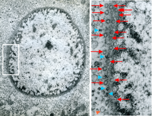
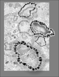
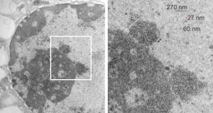
Hepatocyte from a 14CoS -/- mouse (homozygous for a 3800kb deletion on chromosome)that has spent 360 days being rescued from certain demise by NTBC has little condensed chromatin, very round and active looking nucleus and literally thousands of nuclear pores, more than I remember seeing in the nuclear envelope of any other cell type, and any other experimental condition.  Totally awesome. I would not hazard a guess as to how many are there per micron sq.

The purpose of nuclear pores is of course do provide a mechanism for selective transport of molecules in and out of the nucleus, separating the compartment of the cytoplasm and nucleus.  All produced proteins from the cytoplasm required by the nucleus will be imported through the nuclear pore, and the RNAs, mRNA, tRNA and rRNA, destined to become cytoplasmic machinery, transcribed and modified (post-transcriptional modification) in the nucleus will be exported to the cytoplasm through nuclear pores.

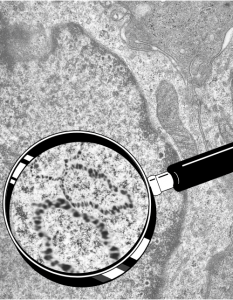
So the electron micrograph below (and inset) show this massive number of nuclear pores, particularly on one side that is cutting through the nuclear membrane at a slightly tangential angle, and also peculiarly, the polysomes (clear spirals of mRNA and rRNA) on the cytoplasmic side of the outer nuclear membrane are just about the only place in the cytoplasm where one finds RER. This vesicular ultrastructure (and particularly the vesicle – within – a vesicle) has been published Deiter et al 14cos, but not the nuclear pore observation. Here is such an hepatocyte nucleus, and inset to show tangentially cut nuclear pores (arrows), and spirals of mRNA with ribosomes (arrowheads). 17709_65053_14CoS-/- 360d w NTBC.

14CoS liver electron microscopy nucleus nuclear pores rescue NTBC


Just a quick comment on nuclear organization

It has been my opinion (based on looking at thousands and thousands of slides and micrographs) that the when the cell undergoes apoptosis or is overly stressed, it shows an amazing nuclear bilateral symmetry.  DIVIDED IN HALF (how does this surprise anyone-yet why can I not find this in the literature)– that is to say that that domains in the nucleus which are beginning to assemble, and/or disassemble in response to the cell death signals, such as the interchromatin granule clusters, and the huge nucleolar structures (fibrillar centers and dense fibrillar components) just seem to appear in light micrographs (where sections are relatively thick compared to those for TEM) in a mirrored symmetry.

I have seen this same phenomenon in white blood cells (particularly bone marrow smears) where the granulocytes were stressed and micronuclei, or bar-like bodies, or segmentation are increased by (in some cases in bone marrow smears from Cyp1A1, Cyp1B1 and Cyp1A2 ko mice and their many variations and exposure toxins like benzo(a)pyrene or TCDD), and in other cases such (in vitro) where RNAi knockdown of the highly conserved anti-apoptotic gene, C9orf82, induced apoptosis and cells showed mirrored nuclear symmetry, and in 14CoS cells that underwent massive synchronized hepatocyte apoptosis.
This is part of the symmetry of the nucleus currently being explored with data mining, fluorescent antibody imaging, TEM-CT, and yes, even the old school “eyeball” approach. Here is an image and diagram of one bone marrow smear where bilateral symmetry of a white cell is very obvious, and though the image is almost a decade old, and I queried the literature at the time I could find no publications on domains,  territories, or mirrored images in apoptotic cells. (BTW the cells below in a bone marrow prep are NOT apoptotic but nicely segmented).
light micrograph bone marrow smear white cell nuclear bilateral symmetry

More microDNAs for cover submission

I must have really made a lot of variations for this cover submission, here is another that I modified this morning.  It is a little confusing, but the cell behind is a has a large nucleus (left middle of the micrograph), which is in distress, with some indications of impending apoptosis, mainly the finely granular chromatin at the inner nuclear membrane, and the prominence of interchromatin granule clusters, and a rather large nucleolus (with prominent fibrillar centers and dense fibrillar components (in preparation for the trip down the apoptotic pathway). And on that image I superimposed a dozen or more of the images of microDNAs that Dr. J.D. Griffith gave me to organize into a cover. So the microDNAs, (of which three are enlarged bursting out in dimensional relief) that are mostly black and grey high contrast, while there are many others lesser in size that are superimposed in relief over the original transmission electron micrograph of the cell.

Electron micrograph of nucleus and overlay with microDNAs

Still working on a cover submission with Dr. J.D. Griffiths microDNA images, superimposed over one of my own electron micrographs of a cell with prominent nucleus and highlighted within that as an inset is an enlargement, over which is superimposed his micrographs of microDNA.

I don’t think this image was selected for submission, but it is pretty nice none-the-less. I am prompted to post these, after looking back over the archives (for other reasons).

DNA and microDNAs

See these images which were submitted, but not accepted for cover illustrations.  Electron microscopy by Dr. J. D. Griffith, and rendition by Dr. Marian L. Miller.  We were working on a cover submission, and those examples below are just a few that were organized, plus the one I liked best shown in a previous post on this  blog here .  The bottom right image I find this morning while googling, skimmed, and referencing some keyword advertizing company…. this makes me furious. The pasted up DNA is middle right, and all these are Dr. Jack D. Griffiths micrographs of microDNAs.  Shame on you skimmers and spammers and hackers.

Interchromatin granule clusters: HeLa cells UV + inhibitor of caspase-1

Interchromatin granules (that is a cluster of granules) of varying size, but mostly rounded areas, are found in the nuclei of mammalian cells. These are clustered in the nucleus, especially in and around apoptosis as seen in the lower magnification electron micrograph on the left below. They are, according to concensus, composed partly of pre-mRNA splicing factors.  They are not static structures, and are active sites of transcription, size can be several microns or hundreds of microns in diameter.  They were found in a study done years ago, to be really prominent in late phases of apoptosis. The image on the right shows particularly well (though the micrograph is not sterling by any means) that they are composed of 20–25 nm granules that are connected like beads on a chain by thin fibrils (also visible).  The red dots in each of these two images (the right image enlarged from red box on the left) are the size of ribosomes, and the bar markers then are 10 times bigger (270 nm).  There are at least 3 other interchromatin granule clusters in the micrograph on the left. That shows the beads in the right hand enlargement to be just around 20-25 nm in diameter (something a little smaller than the accepted middle size of a ribosome (27 nm) (selected from the same micrograph.

Interchromatin granules may be collections of snRNPs and other RNA processing components involved in producing mRNA. These granules are so evenly spaced as to be a marvel.  neg 14138_block 61002 HeLa cells, UV and inhibitor of caspase 1. (BTW, dont ask what the dense areas to the right of the interchromatin granule cluster is…. ha ha)

rRNA transcription just confusing

This is so awesome, just read part of this article by cheutin et al_awesome nucleolar diagram and view the TEMs and diagrams, were that I were beginning a career in TEM tomography, i would be in heaven. But 40 years too early.  So at best, is to take the new information and correlate it with studies from the past, making use of the data that I have and thousands of electron micrographs to play with.

TO begin with, 14CoS nuclei and nucleoli with some bar markers, just getting acquainted with the possibilities for labeling these structures. So here is the image from a previous post, and the segment enlarged with the two cajal bodies, tiny red circle is the size of a ribosome. Nucleolar structures I have not measured yet, but according to the publication by Cheutin et al, linked above, these will be similar and important in the pre-apoptotic hepatocyte nuclei found in this mouse line when they are not rescued by administration of oral 2-(2-nitro-4-trifluoromethyl-benzyol)-1,3-cyclohexanedione (NTBC).

electron micrograph liver 14CoS ko