Monthly Archives: December 2017

Stub tail monkey liver: desmosomal mitochondrial tethering

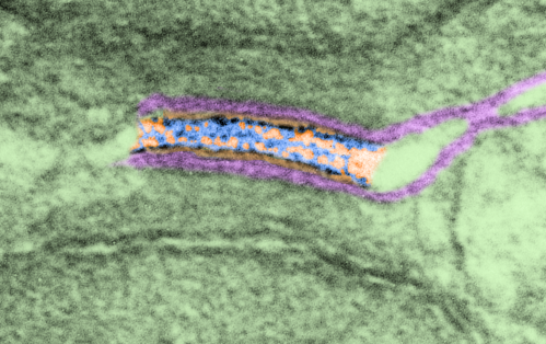
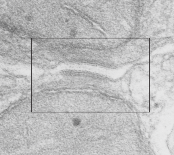
This is just a micrograph to justify the idea that the desmosomal mitochondrial tethering, or junction or des-mites as i have aptly named them (as opposed to pore-mites) for nuclear pore mitochondrial tethering, are pretty universal. Here a single tether is in a stub tail monkey hepatocyte, from tissue taken back in the 1970s while studying the effects of infusing artificial blood emulsions (perfluorochemical based blood substitutes) . This particular monkey (Maccaca speciosa, probably female) did receive two test doses of a perfuorodecaline emulsion, just a tiny amount, 0.05cc/kg of a 10% emulsion PP5ct and 5% F68) (Lee Clark Jr named all his emulsions, this was EM#750428). no perfluorochemical droplets were seen in this hepatocyte. Sac date was 5 10 76.

So desmosomal mitochondrial junctions are here. Mitochondrion is in top part of micrograph, desmosome is attached to the down pointing portion.


More pseudocolored desmosomes and mitochondria – tethered

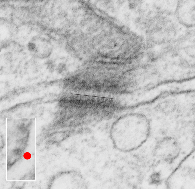
Here is another pseudocolored desmosome with portions of two mitochondria top and bottom parts of this electron micrograph. It was difficult to determine exactly where the plasmalemma from each of these two cells went at the point of the desmosome, but I thought long and hard about which part of the structure they were. It seemed to me that there was a slight electron lucency just under the plasmalemma on each side, therefore this is the way I pseudocolored (with pink) the cell membranes.  The densities within the desmosome itself looks like there are three rows… the central dotted (periodic) line where the cadherin molecules knot together (my guess is this is a totally symmetric arrangement, not at all random like suggested by some) and perhaps another periodicity (well not perhaps…. it is pretty striking) which lies a little separated from the plasmalemma.  I don’t know if any of the models of the cadherins show a “lump” structure before the transmembrane part… ? That will take some searching.

Top image is unretouched transmission electron micrograph of a desmosome, as mentioned, mitochondria portion seen top and bottom.  The box in this image is what is enlarged in the second image. There are two very prominent intramitochondrial granules, especially the one in the mitochondria at the bottom of the micrograph.
This image is from the boxed area above, thus enlarged, pink is the plasmalemma, orange is the area just under the plasmalemma of each cell and into the space of the desmosome. Blue is what I see as the possible densities of cadherin molecules.

In this inset the periodicity of the outer part of the desmosome (probably still cadherin molecules) is a spacing about 1/15nm  not too different from that found in a previous post at something around 1 density for each 13-14nm spacing.  The periodicities on the central dense line of the desmosome in this micrograph might be something around 18nm spacing… I would have preferred if the densities came out one-to-one, but anticipate that in other assessments that it might do just that. But for now, i just count what shows up. 6102_5070_mouse_female_control.

FROZEN? in more ways than one

First the apologetic! Few grew up in an atmosphere more infused with Disney than I did. Southern california born – a father who lived on Mariposa next door to July Garland – steeped in Hollywood culture from afar – a cartoonist in high school – a short-time employee of Disney studios as an “in-between” man (i think my dad wanted more freedom). My aunt Joyce Weil lived next door to Ollie Johnson, long time Disney employee and toy train addict, upon whose backyard miniature steamer?  train I rode as a child – my best friend’s mother painted all the skirts of the fairies (dandelion seeds) in Fantasia – our summer vacations were at Disneyland… my favorite place on earth – and my sister was a cast character at Disneyland for a while – my brother worked marionettes for a disney film – and from time as a toddler to teen years I dreamed of working at disneyland painting the pretty parts of Fantasyland.  My mind is still full of gingerbread-type ideas. I have made stained glass disney panels for the woman who was the original Mickey, and is still Minnie at disneyland and did Pete’s Dragon for one of the lighting engineers. There have been countless family gatherings under the disneyland christmas tree – though now a gathering would be without those who have departed from our presence, but would be filled with new little faces. Basically I wanted to live at Disneyland. I am, because of this upbringing (or at least modified by it) an incurable romantic, waiting still for a prince charming to find me, and to live happily ever after in a fairy-like castle.

The impact of fantasy – on our lives, and perception of life? How good is it, how damaging. I wont weep that my dreams were never fulfilled…. or even suggest that at a wise and sage 74 and having been a scientist for 40 years that that might qualify me to be a consultant on their “imaginarium” team, for biology, histology, and physiology which I would do for free… This wont happen either.

What i will comment on is the impact of that environment had on an already slightly “fantastical” (haha) mind. I wept watching the TV version of Frozen last night. In some way it touched me really deeply, because 1) of the sister thing and the separation (in distance) between me and my two sisters, 2) because of the struggle to understand that ultimately “love” conquers all and 3) because in some ways a profession (mine in science) reinforced the separation that i feel from the fantasy/love/happily ever after part of my brain which I had hoped would be the main path of my life.
My prayers to all of you who have not yet found the “anna” in your hearts, but are like “elsa”- afraid, reserved, frozen.  I don’t know if i would have ever found the “anna” in my own life had i worked in the field that i dreamed of as a child…  maybe, maybe not, I pass no judgement on the impact of fantasy on the reality we all must live with, especially the impact on the young, and young at heart. Reality ≠ Fantasy = Disney.
Desmosome-mitochondrial tethering: just what I see

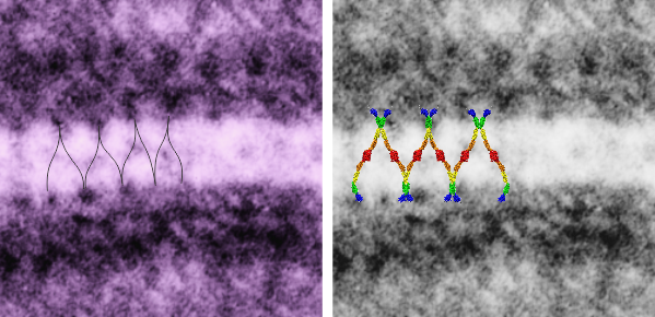
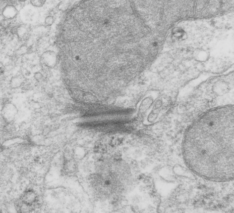
I found this particular electron micrograph which has two mitochondria and a desmosome in pretty good orientation to look at any periodicities or patterning in the substructure. It seemed to me that the central line of the desmosome, and the cadherin molecules which create it and the links to the plasmalemma (which have been modeled with molecular models) could fit best as a “sprint” type association.  Those of you who are old enough to remember the plastic hair combs that looked like wishbones arranged in parallel that one could use to sort of tie up a pony tail, will recognize this flexible structure.  I found, or should say “think i found” a similar type of density in the cadherin molecules in the center of this desmosome (diagrammed as that side by side “wish bone” array.  Interestingly, cadherins were mentioned to one group to come in a dimer which would very easily form this springy wish bone structure….  and listed the “half wish bone” as one option as a molecular model of the desmosomal cadherins. One of those articles has a great 3D image which doesn’t look exactly like what i see, but is close found here    – and another is this title Studer, Daniel & M Humbel, Bruno & Chiquet, Matthias. (2008). Electron microscopy of high pressure frozen samples: Bridging the gap between cellular ultrastructure and atomic resolution. Histochemistry and cell biology. 130. 877-89. 10.1007/s00418-008-0500-1.
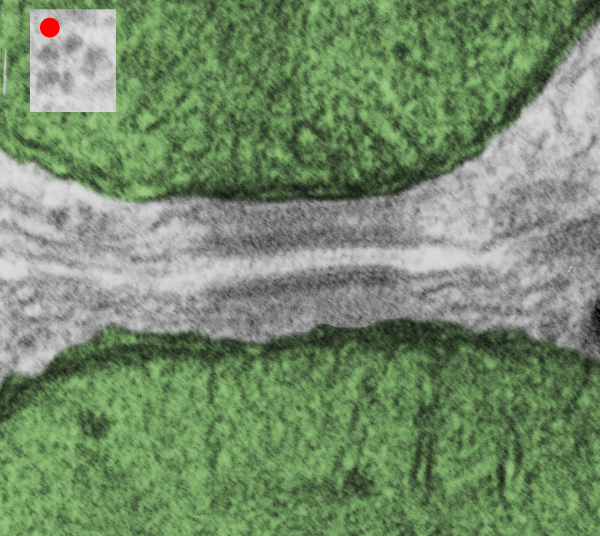
. I actually don’t like the idea of calling the arrangement “untangling desmosomal junctions with knots” as i think if they were knots….there would be no perfectly wonderful order that can easily be seen with ordinary transmission electron microscopy. It is also very appealing to have flexibility within these junctions as the wishbone (wish bone) alignment would afford…. that would be just really fun. electron microscopy mitochondria desmosome junctionSo here is the same electron micrograph as yesterdays post but the mitochondria are actually fuzzed green in a separate layer.  Red dot=a ribosome, approximately 27 nm in diameter. The absolutely regular arrangement of cadherins is seen in the middle of the desmosome…. no knots, no tangles, no mess….  just regularly spaced. This micrograph is not retouched to emphasize anything…. it is just the way it looks on the negative. The plasmalemma of the two cells is not in exact cross section and so it is fuzzed. The span between densities both on the plasmalemmal sides and the center densities of this desmosome work out to be about 1/13-14nm.  I have not put that together with any models yet and in the image below just sized the molecular structure according to the TEM, not using the actual size (just using the shape).

electron microscopy mitochondria desmosome junction
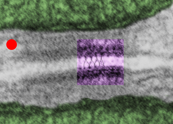
So in the second figure you can see an area that i enlarged, cut and pasted in photoshop, and enhanced in contrast and colored purple  and ADDED what i think are the lines of the wishbone arrangements of the cadherins.  Densities (increased in contrast here) work out to about 1 for every 13-14nm.

Into two enlargements (one the purple box above and another with the molecular model of a cadherin dimer, copied and mirrorred, fit really nicely into what might be a flexible, stretchy springy type portion of the desmosome.  Just a thought….  ??

electron microscopy mitochondria desmosome junction
6110_5080_mouse_f_liver_36,200x_4x


Cadherin molecules overlying a desomosome – mitochondrial tethering

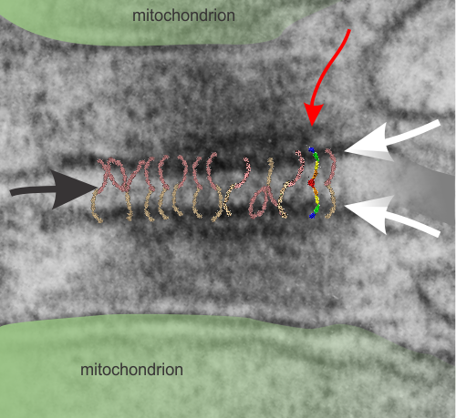
I don’t know much about desmosomes, but they do make unique junctions with nuclear pore intermediate filaments and also very definite connections with mitochondria. The mitochondria – intermediate filament connections with desmosomes show a couple nice ultrastructural changes from the routine: 1) the mitochondrial outer membrane is flattened, or straightened in the area of connection with the intermediate filaments over the desmosome, and it is also a little darker. and 2) the mitochondrial body itself is drawn towards (at least that is the way it looks… as if there was a dragging force) the desmosomal intermediate filaments. The central line of the desmosome is not always visible when i see these tethers (often with two mitochondria in adjacent cells and a single desmosome, but there are molecular models of the cadherins (several) that I cut pasted masked and reduced to fit in the appropriate intercellular space where they would appear. It is a good lesson is relative “size” and “shape”.

Micrograph: Pale green semitransparent mask is over two mitochondria, one each in two adjacent cells. Red arrow points to the inner dense layer (maybe i will add the known proteins there “in scale), the white arrows point to the plasmalemma of the two separate cells, the black arrow points to the center dense “knotted” region, (maybe i will measure distances between visible knots) and the molecules (several taken from the internet, look similar and were pasted into the intercellular region (see demarcation from right hand side transparent grey.

Triple desmosomal mitochondrial tethers

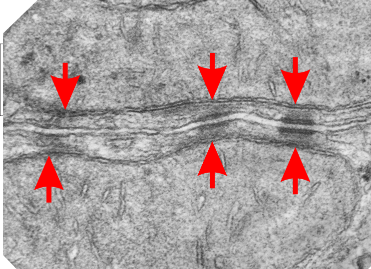
Both sides of two adjacent hepatocytes show two mitochondria each linking to three desmosomes.  Nice to see such symmetry and the implications are quite amazing, in that whatever is happening at the plasmalemmae of these cells is happening “together”, really nice intracellular communication.  Red arrows point to the inner mitochondrial membrane, just above where the intermediate filaments of the desmosomes approach the outer mitochondrial membrane.  I wish i could say that I saw some cross sections of intermediate filaments here, but i didn’t.  The double desmosomal mitochondrial junction on the left is more tangential than the one on the right.  The pair in the middle is intermediate.

three desmosomes two mitochondria six mitochondrial-desmosomal junctions

Intermediate filaments tethering desmosomes and mitochondria

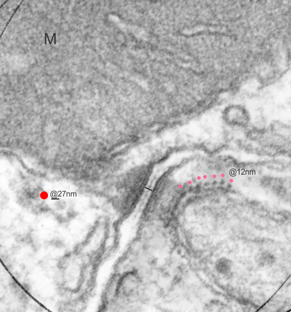
I have mentioned this phenomenon before but i keep finding images which speak to the prevalence of mitochondria (in this image two mitochondria are tethered to a single desmosome, one each in two adjacent cells). in addition to the fun double tether here, look at the “spots” which round the corner of the smaller mitochondrion…. tiny dots of what I measured as about 12nm, but the intermediate filament on cross section is 8-10nm, therefore my guess is that these wre cross sections of intermediate filaments. I did enhance these dots in any way except to change the contrast of the lower half of the image using photoshop.  The dots ” cross section of evenly spaced intermediate filaments” are very prominent on their own.  Pink dots are proximate to the filament x-sections, the red dot is an approximate ribosomal diameter.  30nm bar in middle is about what the desmosome measures, oriented perpendicular to the two plasmalemmae. N 6105 Block 5080 Mouse, female, orig mag 36000

Chromatin at the inner nuclear membrane

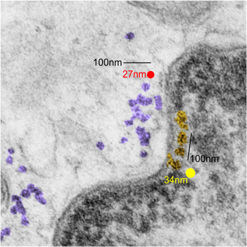
There is a lot of organization here, about which I know almost nothing, but it is fun to look at old micrographs and see pattern.  This is only a view of a portion of a nucleus which has some very nicely defined chromatin (right side of the image, overlay is yellow, which measures not that different than a ribosome (middle and left portion of image, pseudocolored purple, and red dot, taken to be about 27nm) of which there are several in the nearby cytoplasm. Bar=approximately 100nm, and the spacing between nuclear chromatin bodies is less than 3/100nm.

RNP along the inner nuclear membrane

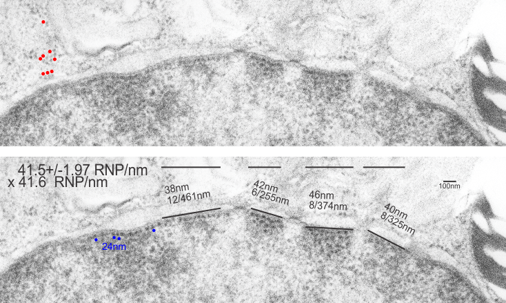
When i encounter electron micrographs where there is such obvious order i just marvel at the detail and complexity of life. Here on the inner nuclear membrane of an alveolar type II cell, in between the nuclear pores, there are little RNP particles, neatly and tidily spaced at about 41 per 100nm, and at about 24nm diameter (slightly smaller than the ribosomes from this same micrograph used at a measure of 27nm diameter. These are organizationally (that is, the RNP and inner nuclear membrane, and the ribosomes on the RER membrane) and with such similar but not identical sizes, shapes and arrangements,  that it becomes almost silly not to see an evolutionary structural relationship.

Picture on top has NOT been altered, but the identical picture on the bottom has had the distinct areas of RNP burned using photoshop just to show you what i see. Red circles are ribosome size, relative to the enlargement of the images (taken at 27nm) and blue circles are RNP granules, which are apparently closer to 24nm diameter. Point here is the rigidity of the inner nuclear membrane, and flexing of the outer nuclear membrane at sites where the ribosomes on the outer nuclear membrane are actively producing protein.

Four adjscent inter-nuclear pore distances have been measured and RNP counted. Mean+/-SEM is given at 40.5 ribosomes/100nm  and all ribosomes together with all nm is 40.6. A tangential area of the inner nuclear membrane and many RNP forming a grid like network is seen about midway-top of both micrographs, but accentuated in the lower micrograph.