Monthly Archives: January 2018

Different intercellular joints?

Weld (stronger than rivet)*; light in weight; can be made with access to one side only; making and breaking requires little energy= tight junction
Rivet (loss in strength); some flexibility; heavier; requires access to both sides (2 cells); making and breaking requires energy; = desmosome
*http://www.1920-30.com/architecture/rivets-welding.html : this source says there is no loss of strength with a weld, but particularly, when i viewed micrographs of spot welds, sometimes the sheets of metal look thinner and the texture of the metal has changed under the electrically produced weld.

The desmosome is not really a butt joint, since the center plate in a butt joint is the intercellular space and there is no edge to be joined in the respective adjacent cells. Maybe it could be considered more like a double-rivet lap joint. Or a double-rivet double strap joint.

Environmental impact on desmosomal joints are: tack ( how easily a bond is made and broken  ); peel (the force needed to break the bond between two cells – including peel angle and direction); shear (sliding between surfaces), and then there is the list of molecular forces (Ions, enzymes, water, heat and energy).

Here is an interesting quote from label makers, “Non-carboxylated emulsions tend not to build adhesion over time, an important attribute in removables. Tg influences pliability, especially at low temperatures. (Think labels applied in the freezer section). Gel content is a measure of cross-linking within a styrene-butadiene emulsion polymer. Less cross-linking results in higher tack, but more cross-linking provides higher shear. If necessary, a tackifier can be added to the emulsion to raise tack even more.” and one wonders how this polymer science stuff relates to the polymers in desmosomes.

The desmosome, the adherens junctions — both have attributes, there is a  trade-off from one type to the other, for different jobs and  modifications  within each category.

Shear testing for desmosomes, as layered structures,  disruption along the plane of the cell membranes. I guess a desmosome can be called a viscous adhesive.  haha. or a velcro spot.

Desmosomes: ductile, double-sided, shear load rivets

Desmosomes are kind of like Oscar rivets, and are blind perhaps, or double-sided. the latter have splits (the desmogleins and desmocollins) along the shafts. These splits cause act like flares and join in the central dense line (periodicitities) of the desmosome. The plakoglobins and plakophilins form the “head” of the rivet along with desmoplakin, and these rivets are unlike most rivets found for metal and wood work, because they are flexible, and have a  “spring-hinge” at their head, the desmoplakin-intermediate filaments junction (which i believe is perpendicular not parallel (as most of the diagrams show), which can flex and bend and allow for motion of the desmosome as it gathers together as weld between two cells.

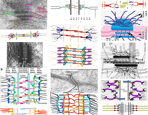
Below is a set of diagrams taken freely from the internet from publications on desmosomes (including my own). It can be seen that the majority of diagrams don’t really “see” what the microscopist sees as the course that intermediate filaments take when they are adjacent to desmoplakin.  The diagrams on the lower left, and all those in the center and the lower right have intermediate filaments coming from the cytoplasm and making hairpin turns at the desmoplakin molecules…. However, the micrograph on the upper left, and the diagram in the upper right have the intermediate filaments which actually appear to arc ever so slightly, to be NOT forming hairpin turns to desmoplakin, but gently curving perpendicularly to the desmoplakins.

It seems more likely that the type of shear stress (the connection between desmoplakin and the intermediate filaments perpendicular to one another) would be more advantageous at binding two cells together than the diagrams below (with hairpin turns) which suggest that the connections between intermediate filaments and desmoplakin are parallel, thus involved in tension stress.  The former intuitively suggests more efficient shock absorption over a smaller distance than the latter, and actually looks more like the actual anatomy than the latter.

The disparities among the images from electron microscopy and the diagrams from the literature are the reason that I began to try to think this through. Intermediate filaments are seen in micrographs that look perpendicular to desmoplakin (like what is seen in the micrograph in the upper left part of the diagram as brown lines), or as 10nm round dots (if they are cross sectioned).  Any ‘apparent’ long intermediate filaments parallel to desmoplakin (as seen in the bottom center electron micrograph) can almost always be explained as tangential cuts.

This arrangement is most clearly demonstrated when intermediate filaments form a flat perpendicular band between the desmoplakin molecules of the desmosome and the mitochondrion when the desmosome and the mitochondrion are tethered together.
diagrams of desmosomes

Oh brother!

As regards a study by several researchers that found that the ability of children to exhibit executive function improved when they were empowered by how they were dressed (this time in super hero costumes) which allowed them to focus on work (avoid temptation of playing on their ipads) for longer periods of time.

Then come some editorial comments…. the negative comments i read all came from men….. i remember when (40 years ago) someone got an NIH grant to study bonding between mother and newborn and they were ridiculed and mocked and became the laughing stock of science….how sadly misguided then, and how sadly misguided now….

Only idiots would negatively comment on a fundamental maturation principle, as simple but as important as delayed gratification as being unimportant research. As it was suggested thousands of years ago, delayed gratification (sanctification) of the everyday carnal (fleshly) experience is what adds superior meaning to life, and enhances frontal lobe function. (And that, for those of you who don’t know it, is the difference between human animals and animal animals.)

The world currently thinks instant gratification is their due…. like trump… who takes what he covets, and has no executive function whatever (lest a trump advocate reads this, please note that executive function here does not refer to the office of the presidency, but to frontal lobe governance)(and lest a person in public governance reads this, governance here does not refer to running of the office of the mayor or the governor)

Desmosome dimensions relative to adjacent membranes and mitochondria

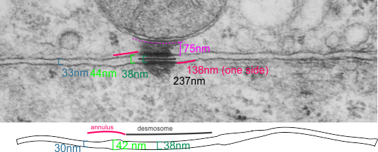
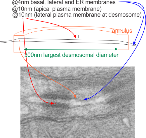
It seems to me that there is a pattern of thickness  changes(width or height if you wish… because of the orientation of the diagrams below) and the rigidity of the plasmalemma inherent in the desmosome (likely due to the transmembrane parts of desmoglein and desmocollin ) and that rigidity includes a specific intercellular space dimension. I have seen this published at about 34-38 nm (that is, from the inner leaflet of the plasmalemma of one cell to the inner leaflet of the plasmalemma of the adjacent cell) to be something on the order of 38 nm.  Using that dimension, if i measure from inside plasmalemma of one cell to the adjacent cell and compare that to the width from the same places in the 200 nm ring or annulus around the desmosome there is going to be a change in the dimensions (which i could measure as just a few nm greater than that within the area of the desmosome proper. And, then a reduction in the intercellular width (per the more routine proximities of two cells) which becomes something like 40 nm.

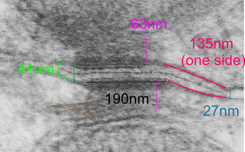
Here is an article which suggests some dimensions for the desmosome, but does not address adjacent variations in the plasmalemmae including the anulus.  You can compare with two images and measurements below. Keep in mind that the two images below are not derived completely randomly, as I picked two which had substructure which was clear enough to measure.  The annulus of these two desmosomes is indistinct, and longer than that seen in other desmosomes. This likely relates to the plane of section on a perpendicular axis of a round desmosome and surrounding annulus (the latter being increasingly seen as one sections nearer the periphery of the actual desmosome).

top diagram shows a length of two adjacent plasmalemma, measurements relative to that image, bottom one is a different desmosome, notice brown lines for intermediate filaments, black line for length of the desmosomal spot, dotted lines at periodicities of the bridges between desmocollins and desmogleins, outer dense line with dots, periodicities of the extracellular membrane anchor, pink bracket, thickness of the desmosomal intracellular elements, which includes proteins of the intracellular anchor (desmocollins and desmogleins) plakoglobin, plakophilin, desmoplakin and intermediate filaments (visible at the desmosomal-mitochondrial tether on the bottom more clearly than the desmosomal-mitochondrial tether seen at the top of the lower micrograph.  While a big deal is made of the inner and outer dense plaques of the intracellular part of the desmosome, the lower portion of the lower micrograph doesn’t make that case.  Were the micrograph sectioned end on to the intermediate filaments below, there might be a more visible inner dense plaque.  The outer dense plaque (plakophilins, plakoglobin portion and desmoplakin proteins) is well defined. NB, there is a “flatness” or “rigidity to the outer mitochondrial membrane where the intermediate filaments lie beside it… i hope to search for proteins that might be involved in the linking of mitochondria and intermediate filaments.


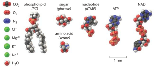
Biology by the Numbers: great idea

I have found this website (i think i mentioned it before) but I really love it. Most textbooks (or i should rephrase this to be, textbooks of my era) don’t think about providing relative “size” and “distribution” to their descriptions biology, cells, organelles, etc. Somehow those relative size tidbits are what help me remember and understand what I read.  So this kind of a chart for me is awesome.  I laud the editors of this book…   I WOULD LOVE TO HAVE a resource where they just had allllll the known protein structures that I could look at and superimpose where they belong over my electron micrographs.  ha ha. Someday, someone will be able to do that.

Desmosome dimensions

I found a cryo-EM paper of desmosomes that was pretty nice.  Well desmosomes in the lower portion of the epidemis might be like desmosomes in liver, and it is very clear from this paper and others that the desmosome is adaptable, become different things in different cell types and tissue types. So this is a general description, but the basics are present in desmosomes of the liver.  The measurements they show do NOT exactly fit what is stated for the “viable epidermis  desmosome this publication states, but they are pretty close to those that I have re-measured just from their image, their bar marker, and my own repeated measurements from densities to densities.

The measurements I came up with are posted on the micrograph. The baseline measure came from the micron marker given for their own image. The measurements I made are for inner lamina to inner lamina of the two plasmalemmas; outer lamina to outer lamina for the two plasmalemmas (extracellular width); the pretty lucent area just extracellularly to each plasmalemma, the length and dimension between the desmoglein and desmocollin molecules (cant tell which is which) and also the “v” type structure, alternating and overlapping as the center dense line of the desmosome (in the extracellular space).

I thought it was interesting that one researcher called this intercellular linear pattern random, while another called the pattern linear…. in fact i think they both missed the pattern and I have outlined the cadherin rhythm in green. The distance between the repeat portions (ectodomains) of the latter looks maybe to be close to linear, or slightly curved, and the N terminals (to my way of thinking) create a picket fence, or zipper kind of pattern.

The micrograph I used for these measurements came from a paper by Ashraf Al-Amoudi, Jacques Dubochet, and Lars Norlen which is available online. Their bar marker is at the top (50nm) and all other measurements were made from this distance.
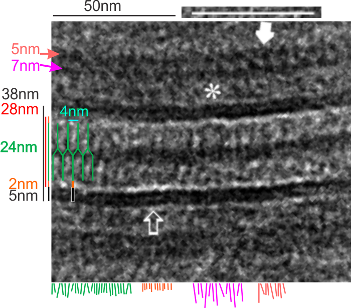
5nm approximate thickness of the cell membrane (one on each side of the desmosome shown here)
38nm from plasmallemma through to plasmalemma of second cell (THEIR MEASUREMENTS LIKELY TO THE CENTER OF THE TRILAMINAR MEMBRANE were about 33nm — pretty close)
28nm extracellular space (THEY DIDN”T MEASURE)
24nm is the @ height of the repeating units and N terminals of the desmoglein and desmocollin molecules (two molecules stacked=height (vertical dimension in this micrograph) (THEY DIDNT MEASURE). It is marked as the green stretchy-wire lines (likely a significant configuration for movement of cells that allows for some “give and take before break” which would not be surprising, but actually be awesome) and was easily drawn over many portions of many micrographs (this, and others of my own that i have posted before) .
4nm spacing between the repeating but alternating units of the desmocollins and desmogleins (THEY THOUGHT THAT THE VERTICAL LINES WERE 5nm APART, I think more like 4nm apart and staggered).
2nm orange lines, the lucent region just before the transmembrane segments of the desmocollins and desmogleins.
7nm pink arrow is a measured periodicity of one band of densities in the inner plaque, and the orange arrow represents measurements from adjacent periodicities.  To me they didn’t look the same… the latter perhaps being further spaced and still alternating. White asterisk is from the original micrograph in the Al-Almondi paper, outer white arrow points to the same group of proteins as my orante arrow labeled with 5nm distances. Their white outline arrow points to a lucent area that i did not measure which is just intracellular to the inner leaflet of the plasmalemma.
My measurement lines are shown below the micrograph. Green: (i should have made black since they were measures of plasmalemmal thicknesses).

In liver, the intercellular space between hepatocytes is actually greater than the extracellular space of the desmosome filled with the cadherins….  which really is a very rigorously attached and spaced area.

One other difference in the way that Al Almoudi describes the “inner dense plaque” (most medial area of the intracellular desmosome structure) as having a single periodicity…. but when i counted distances between periodicities they actually were statistically different (p=0.03) with the most medial band of periodicities being further apart than those just adjacent and closer to the plasmalemma (but both in the inner dense plaque.

If i would going to hazard a guess on the shape of the molecules that make up the intercellular space of a desmosome i would have one which has a “blump” before the transmembrane domain, a blump at the N terminal (which is an obvious feature as the central periodic line in the desmosome).  The transmembrane part would be quite thin since there appears to be a lucent line just on the outside of the plasmalemma of the two adjacent cells, and before the linear densities are obvious. The current protein databases make an extracellular domain 3-D molecule for desmocollin and desmoglein have a good chance of being fit to the densities found in electron micrographs.
Randomly arranged: not likely

Looking at a summary of desmosomes (so called macula adherens — spots of adhesion) one quote was “randomly arranged” which probably is naive. I find desmosomes (in the only tissue where I have looked seriously… hepatocytes, from several species) to something around a half to a micron, when found cut in the widest diameter. (that diameter There are many instances where the distance between the desmosome and the microvilli of the bile canalicululi is occupied by an adherens junction and a tight junction (pretty much common to epithelia). This is the point where i would have issue with the comment “randomly arranged”, in my experience (which is only limited by my age, occupation and brain) suggests that “very little in biology is random”.  (check out this site for bio-numbers) that says the trilaminar membrane is between 4 and 10nm wide.

It is pretty clear that the intercellular space of the desmosome is constant “height” (approx 10nm) as opposed to the dimension across the diameter (approx 300nm at the widest point) which varies depending upon whether the cut is at an equator or tangential to the desmosomal spot.
It is pretty clear that the plasma membrane within the diameter of the desmosomal proteins is also rigid (owing to some kind of stabilization perhaps because of the transmembrane domains of the cadherin proteins that make up the intercellular portion of the desmosome.
It is also pretty clear that whatever is present in the outer mitochondrial membrane (some kind of link and presence of some type of intermediate filaments) that tether the mitochondrion to the plaque proteins of the desmosome also creates a rigid appearance to that membrane.

Desmosomal-mitochondrial tethers (4) near bile canaliculus

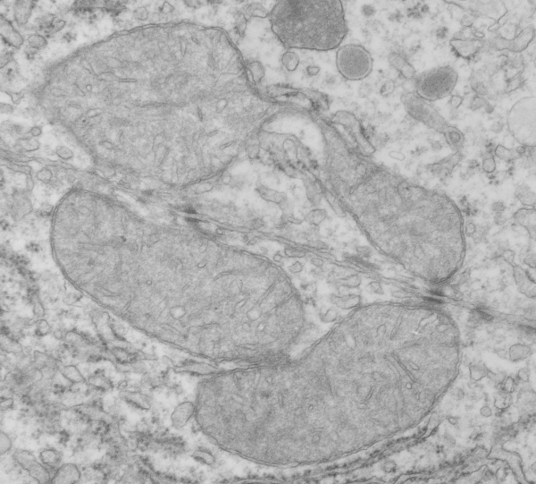
THis Electron micrograph has at least two mitochondria that are attached by intermediate filaments to desomosomal junctions. Adjacent hepatocytes from a rat, which was exposed to carbon monoxide while pregnant with pups. I have to presume this biopsy was taken at the time pups were delivered, around GD 21.  Incidentally there are some funny mitochondrial shapes here, and lots of vesicles,  and peroxisosomes. I do not remember if any morphometry was performed on these liver samples.