Category Archives: Electron microgaphs of lung

Various species of mammal and maybe a non mammal now and then have been obtained and examine using routine transmission electron microscopy. These are summary images.

MVB/LE/PFC: multivesicular bodies, late endosomes, perfluorochemical inclusions

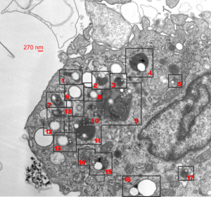
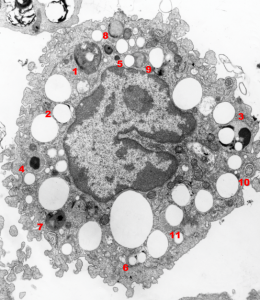
Alveolar macrophage from a mouse lung, the latter having been subjected to complete submersion in E2 perfluorochemical for three hours, and allowed to recover for 48 hours.

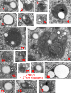
A single macrophage has almost all the PFC inclusions with high concentrations of enzymes, making them appear to have black “caps” in some cases, and in others, mostly enzymes, showing the tiny E2 particles and larger particles (does anyone know if this is Oswald ripening…. it seems as if the particles are doing two things, getting smaller and coalescing…??). The variety of multivesicular bodies here is quite amazing, with some having other structures included, some being filamentous, mostly being very very electron dense.  Each of the MVB/LE with PFC inclusions (i did not tag them all, it was too cluttered) have a bounding box indicated on the unretouched first image,  they are all enlarged and collected in a gallery below (numbered so you can find them).  Compare the variety here with three other posts on MVB/LE/PFC.

MVB/LE/PFC in macrophages can be other than rounded

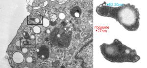
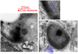
Multivesicular bodies in alveolar macrophages are generally roundish, not always perfectly round, but more round than elongated.  Here are two multivesicular bodies-late endosomes with inclusions which are presumed to be E2 left over from liquid breathing (MVB/LE/PFC) which have lots of protein enzyme content (way more in a volume ratio than E2) and have an elongated shape.  There is a large reaction to the E2 droplets in terms of enzyme production.  So the ratio of perfluorochemical to protein in the lysosomal structures might give a clue as to the characteristics of “export” or “offgassing” of these liquids from the body. The size of the smallest E2 particle in these micrographs is slightly larger in diameter than a ribosome (taken from the same micrograph and enlarged similarly (two insets at right are from the respective boxes top and bottom in the portion of the alveolar macrophage, and there are many MVB, different sizes and many E2 droplets with different amounts of enzyme.  I would guess that the smaller droplets are more enzyme packed than the larger droplets — equating to residence time within the macrophage cytoplasm???

MVB/LE/PFC bodies within alveolar macrophages of liquid-breathed mice

In this case the liquid which the mouse breathed for 3 hours and allowed to recover for 17 days was E2.  Thank goodness for good record keeping otherwise I would have forgotten what E2 stood for. Here is what the notations say (from back when Leland Clark Jr. was keeping tabs on all documents and publications.  E2 (made by Du Pont) O2 solubility ml/100ml at 25oC 55.7, vapor pressure 56mmHg at 37.5oC, viscosity 0.6 at 25oC, boiling point at 104.4oC, density gm/ml 1.656 at 25oC, surface tension 12.9 dynes/cm at 25oC.E2 liquid for breathing perfluorochemical molecular
There were a bunch of MVB/LE/PFC organelles in these macrophages that I began to look more closely at the parallel leaflet patterns that were showing up at higher magnification.

Altogether there were 29 mice that breathed E2, it much have been a satisfactory fluid or there would not be a dozen or more mice that survived from 5 days to 449 days after 3 hr of breathing E2.

The electron micrograph below was one such body, I don’t think it could clearly be labeled a multivesicular body or a late endosome, but there are E2 droplets involved in its organization.  I don’t believe there is any outer nuclear membrane association, but there are membranes with different distances apart which are clearly organized (actually a parallel disorganization – if such a thing can (and it does visually here) exist).  So I sensed that the distance between the leaflets of these membranes was different within the MVB/LE/PFC thingie and in the membranes arranged just outside the MVB/LE/PFC structure and they were different. The latter look to be about separated at 22nm n=28, x=0.053+0.0014  (mm, on the micrograph translated to close to 22nm in actuality) , and I also measured couple of dozen distances between parallel pairs within the MVB/LE/PFC structure itself and it was closer to 28nm (n=31). When the long string of measurements were subjected to a t-test (remember this is an N of ONE macrophage (there will be others) so it is not really saying much) there was an highly significant (p=0.0001) difference between the mean and SEM of the two (color coded) groups. Pictures below show how the lines and data shaped up. White dotted line outlines the two areas sampled for leaflet separation.

Figure below that shows actual sites of measurement and place of measurement for each group.

liquid breathing electron microscopy mouse lung E2

liquid breathing electron microscopy mouse lung E2In another macrophage –another micrograph — another block same E2 treatment and duration, a similar type of inclusion is seen, but this one did not display the linear qualities of the MVB/LE/PFC as above.  There are two droplets of presumptive E2 in this image, they are middle right and upper left.  Whether the lucent area in this inclusion is E2 is anybody’s guess.
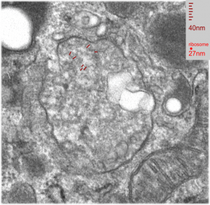
Mouse, liquid breathing E2: macrophage and lysosomes

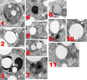
Here is a gallery of the types (big variation) of lysosomes that I found in a 1000A thick section of a single alveolar macrophage from a mouse that had breathed liquid E2 for 3 hours and then was allowed to recover for 17 days before euthanasia.

The only duplicate in style was found in the those MVB/LE/PFC (multivesicular body/late endosome/perfluorocarbon particle) that displayed a small very dense black cap, likely to be the distribution of the proteins.  Sometimes these contained one or two small droplets of E2 (6) or the densities were found at the margin of one large droplet of E2 (2) and even these have different morphologies. A less dense background and divided density contents of some MVB/LE/PFC bodies were NOT arranged with enzymes (proteins?) at the periphery only (3, 5, 7-9) and some polarity to the distribution of the darkest portion (more enzymes) of the MVB/LE/PFC. The lysosome 3 is quite interesting, with three definite PFC droplets and a very marked separation of a less electron dense background and dense compartment (presumably enzymes, maybe portions of some other proteins or even surfactant lipids.  Lipids would be a good guess since they are so darkly stained with the osmium tetroxide). The difference in the size of these droplets is interesting as well, since it seems that the smaller droplets (and maybe they shrink to not detectable with TEM) could be the ones which escape the cell and are offgassed in the lung.  I am just linking the complicated stuff on wikipedia about this, but you can be sure that the temperature difference between deep tissue and lung, and size differences in droplets makes a huge difference in vapor pressure…. someone else can figure this out).  Figure 5 below also shows the tiniest E2 droplets… actually interestingly also noted that the droplets are for the most part found on the periphery of the bounding cytoplasm (likely membrane bound, but perhaps the PFC just interfaces with adjacent proteins in a manner that looks membrane bound (that said for those droplets that do not LOOK to be inclusions in MVB/LE/PFC structures… but then, my guess is that they are all membrane bound having been taken in endocytically.

So the variation is huge, some, like image 1, look like there are lamellar body proteins resembling surfactant and lipids.

There are other MVB/LE/PFC structures which show a distinct lamellar nature with the distance from one dense line to the other is about about 32nm (see below). These are from 1, 9, and one not shown in the gallery above, but just cut off on the corner if image 1, but present in the whole alveolar macrophage at the bottom of this post.

Chromatin exclusion zone and nuclear pore distances apart

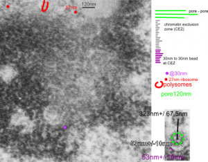
The nuclear pores are really abundant around the bounding membrane of the nucleus, especially in fetal liver cells, and other metabolically active cells. This begs the question as to whether nuclear pores can be measured in order to predict their frequency, and the width of the chromatin exclusion zone around them.  The diameter of nearby ribosomes, taken at 27nm, is used as a standard, as is the diameter of the chromatin beads just inside the inner nuclear membrane, just slightly larger than the ribosome. The inter-bead distance (aka the distance between-chromatin-beads at the margin of the chromatin exclusion zone) and distance from nuclear pore to this string of chromatin “beads”  of the condensed chromatin beside it has been made, along between pore distances This being an alveolar type II cell instead of an hepatocyte, didn’t really define any great differences in these three measure compared with hepatocytes.  10291_18339_ferret_1_alveolar type II cell. Mean distance between nuclear pores in 6 samples so far is approximately 332nm+/-30nm (SEM)

transmission electron microscopy alveolar type II cell ferret tangential nuclear pores

More nuclear pores, just randomly collected various cell types

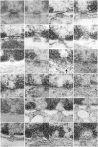
More nuclear pores, just randomly collected various cell types. The criterion that I used was simply the presence of vertical filaments (either part of the nuclear pore basket on the nuclear side or the filaments in the cytoplasm on that side. These come from lung alveolar type II cells, from hepatocytes, and from CoS14 ko mice, rescued and non-rescued, and probably a couple from Gclc conditional KO mice. These are just to give the limit of what effects random sectioning through a block of tissue can do to a round or discoid object. These pores are cut perpendicular (side views), and chromatin is always on top, cytoplasm is always below.

Wait just a minute! is this amoeba lipid or intra-alveolar surfactant?

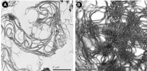
Wait just a minute! is this amoeba lipid or intra-alveolar surfactant? ha ha… I was googling liver toxicity and up came this article (from an image I selected to view the page) and i thought to myself… this could be intra-alveolar surfactant in the alveolar space (left) and tubular myelin in the alveolar space just organized in the center of the image on the right.  This just goes to show us that some principles and some patterns of molecules and physics are going to show up in surprising places.

The article itself was on 3D representations of some molecular organizations in cells, it was interesting as well and linked here.

Spit wad

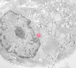
Sorry for being silly, but this alveolar type II cell just made me laugh, as I was dodging and burning to even up the micrograph I saw this cute little nuclear “face” and a little lamellar body spit-wad being sent out to the cytoplasm.alveolar type II cell electron micrograph joke

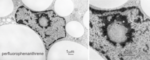
Perfluorophenanthrene in baboon lung

I don’t have a lot of data about the inclusions in this “apparent” alveolar macrophage (my best guess) but the round droplets seen at the left are perfluorochemical emulsion particles which is typical of what was seen in many species after intravenous infusion of such emulsions during studies on blood substitutes during the 1970s.  This nucloeus caught my attention because I though perhaps I was going to find more “ring or donut” shaped nucleoli, but on closer look this area within the nucleus is infact a slightly curved invagination certainly caused by the pressure of one a smallish perfluorophenanthrene droplet on the side of the nucleus just below the section. Inset to right shows the nuclear pores which have been tantentially sectioned confirming that it is not nucleolus.

The range in size of perfluorocarbon droplets within cells is pretty amazing, and can range from sub-micron size to hundreds of microns in diameter. Size of particles plotted against time were related in some way to the off-gassing of the PFCs.electron micrograph baboon lung perfluorophenanthrene nucleusThe widest medical use for perfluorochemicals today seems to be in ocular procedures for the detached retina (vitreous replacement?).  Not for me. no thanks.  Silicon oil would not make me any happier.