Category Archives: Electron micrographs of liver

Size and location of nucleolar bodies

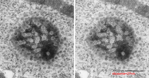
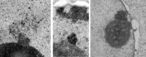
It is remarkable that these little segregated areas of nucleolar constituents look so consistently like “pop ups” that have a predetermined location, density, relative size, and texture.  I don’t believe that it is known what these early blips on the surface of the nucleolus are, (not including the blips that have the substructure notable of cajal bodies) though these may be precursors or similar structures. The banding (linear beads on a string appearance as if wrapped in a ball) in these bodies (by reason of tissue preparation or observer bias –shown in these three micrographs) would not qualify as cajal bodies. So what are they.  The bodies I am speaking of are all pointing upward like little volcanos (especially on the left)  with elements coming or going… whichever haha. Two images on right are slightly more electron dense than the adjacent nucleolar elements.

These images are from scattered micrographs, though two are from 14CoS hepatocytes which had never had or been withdrawn from NTBC.

Heterochromatin exclusion area around the nuclear pore

One cell from the liver of a control (sort of) for the Gclc ko mouse (that is that it had one wild type allele for the gene for turning on albumen synthesis (WC/ii  ) which rescues the would-be Gclc-/- mouse from death. This hepatocyte is not really remarkable to me but it did have this great tangential section of the hepatocyte nucleus and the nuclear pores were just scattered all over the area sectioned making the uniformity of the condensed chromatin areas surrounding the pores very visibly7 consistent.  It was so nicely consistent that I decided to try an afix a casual, but hopefully useful, measurement to that exclusion zone, at least in the this particular animal model.

So, that distance (based on the 27nm size of a mammalian ribosome turns out to be the following:  mean distance, 58nm.  From the original scanned image 58 nm+/- 16nm. That makes the space around the nuclear pore sort of a ring, of about 55 nm. Black line composit measurements, see bottomright. Red dot = one ribosome, electron microscopy nucleolus condensed chromatin exclusion

Nuclear inclusion/invagination? which?

16040 65718 liver 14Cos 24 hr no NTBC, unidentified cell, not likely an hepatocyte, maybe an interstitial cell. Mitochondria in adjacent cells top left and right have condensed mitochondria.  One of the interesting things about the “inclusion” in this nucleus is that it does not resemble any cytoplasmic structure in the cytoplasm of that cell, in fact the cytoplasm of this cell is quite different with lots of vacuoles (lower left) and an ill-defined shape, on one side very scant cytoplasm, which makes it less likely to be an hepatocyte than an interstitial type cell.

Three areas along the margin of the inclusion have been cut and enlarged (denoted by the three boxes in the original micrograph). The inset at the top shows there is really no distinct double membrane typical of the nucleus (inner and outer membranes and nuclear pores) and this might be a tangential cut-artifact. In the inset from the middle left, theer is a band of chromatin along the left border of the inset but again, no double membrane typical of nucleus. The inset from the lower right abuts up against nucleuplas which is very electron lucent, but along this area there is a density to the inclusion which looks like it might be some kind of lamin proteins.

Another great feature is the constriction appearing in lower middle of the inclusion, densities that look like they might have been some kind of “ring” around part of this structure.

Ribosomes (estimated at 27 nm in diameter from the cytoplasm have been enlarged with the insets (also shown in the original).  Bar is 10x the ribosomal diameter at approximately 270nm.

The inclusion/invagination is full of robosomes and RER membranes (I guess that is what they would be named) with an homogeneous appearance and the vesicles or RER appear to be dilated to about the same uniform degree.

There is a nucleolus just above the inclusion. Animals with genotype undergo a pan-apoptosis among hepatocytes within a few days of birth, this doesn’t look like apoptosis yet, and also doesn’t appear to be an hepatocyte.

electron micrograph liver 14Cos ko mouse

Electron micrograph of an hepatocyte (gd 20 fetal liver)

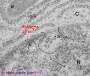
From a study in the 1980s looking at exposure of pregnant dams to CO or dichloromethane by inhalation, here is a kind of “pristine” hepatocyte from ad gd 20 fetal rat. The nuclear pores are numerous but not really that distinct, and the RER is nice and flat but also has a dense content, and the mitochondria have cristi that are not completely flattened and there is not a lot of it, and very little SER that I can find and this cell is adjacent to two hematopoietic cells, no desmosomes between. M=mitochondrion, C=cytoploasmic side, N=nucleoplasmic side arrows point to condensed (closed) and euchromatin (open). Ribosome can generate a relative size comparison for other structures, being about 27 nm in diameter (red dots), the nuclear fibrils just at the nuclear pore (almost dead center in the micrograph and within the black dotted lines) about 30 nm (black) and perichromatin granule in purple. The project, as I recall, did not uncover any differences between the exposed and non exposed animals, and in this case, the hepatocyte looks nice.

Working out dimensions of nuclear and nucleolar components from archived electron micrographs of hepatocytes

It is nice to read that something is measurable, that has a dimension in nm that one can put into some kind of life-perspective. I don’t know how accurate those individuals were in giving a distance measurement to their images are, and I am giving each the benefit of the doubt in terms of attempting to be accurate) i guess they are quite accurate. I know for myself, using three or four different electron microscopes, and various different paper products, films, dryers, etc, that there is shrinkage, stretching, and all sorts of changes that have occurred over time (even 35 years) that affected the dimensions of objects on the micrographs, even though by a very small amount.

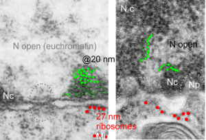
I am using 27nm as an internal control for magnification+enlargement of images. In this case, the 27nm red dot, in all images has been derived from about a dozen ribosomes, and labeled as such. The green dots are about 18-20nm in diameter in these micrographs, and strung as if “beads on a string” (not the beads on a string of nucleosomal DNA, but production of preRNA in the nucleus?) but is it not strange that there is a repeated motif – that is- beads on a string- showing up a lot, both in the nucleoplasm, the nucleolus and the cytoplasm (ribosomes on mRNA) as a kind of “convenient” little pattern.  And who knows where else this pattern might be found). So the green dots show this motif, in a parallel-linear pattern, in the condensed (or closed compartment) chromatin. Both of these examples come from Gclc ko mouse hepatocytes.  Red dots are ribosomes.

Nucleolar ultrastructure: fibrillar center organization

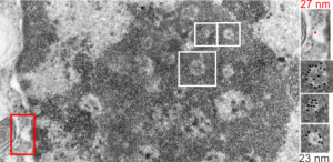
I was looking at this micrograph of a n hepatocyte with the fibrillar centers and dense thick fibrils in their midst, and the granular zone around the outside of this nucleolus. The latter being very punctate in appearance, just really obvious. I measured these once again at about 23 nm in diameter and noticed that around many of the fibrilar centers where was a basket, or ring, or radial distribution of these small punctate appearances.  OK so they are not really punctate, but can appear smeared like fibrils too, though the granular appearance and the nomenclature is well entrenched in the literature, they are just going to be strands not dots.  It could be that these radial dots around the fibrillar centers are bands, or windings at a given distance apart, kind of like fingers of a basket, or more aptly put, like a cage, with wires of 23 nm in diameter.

Electron micrograph here shows a portion of a nucleolus with three fibrillar centers cut out and enlarged (original sites are boxed in white), and a portion of the outer nuclear membrane with ribosomes (for measurement – one ribosome being about 27 nm in diameter, top right image with red dot and text). The cross section of the cage, or basket filaments around the fibrillar centers are shown as black dots.

Estimates of the size of some nucleolar components

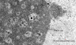
This micrograph, parts of which have been seen before, is an hepatocyte nucleus which is in the early stages of demise (untreated CoS14 ko without rescue). The nucleolus is large, and has two cajal type bodies (one seen here) and fibrillar centers with fairly large densities within (of approximately 90 nm diameter). I have given these sizes (relative to a ribosome size red dot approximately 27 nm (lower right hand side of micrograph labeled as such). The granules within the granular portion of the nucleolus (as well as the banding patterns found on cajal bodies) is something around 23 nm.  What I presume to be perichromatin granules look to be about 50 nm. The large dense bodies within the fibrilar centers are a little unusual.

Desmosomal-mitochondrial associations

The physical proximity of mitochondria to filaments in the cytoplasm has interested me for a long time. Particularly, those associations between mitochondria and the cytokeratin filaments (tonofilaments, intermediate filaments) on the cytoplasmic side of desmosomes and the associations between mitochondria and nuclear pore filaments (those projecting from the cytoplasmic face of the pore into the cytoplasm). Anchoring plaque proteins and central proteins are pretty nice in this electron micrograph which wikipedia describes thusly: “transmembrane linker proteins, called desmoglein and desmocollin, belong to the cadherin family of cell-adhesion molecules bind to plakoglobin and other proteins in the plaques and extend into the intercellular space, where they interact, forming an interlocking network that binds two cells together”.

I have hundreds of examples of these interactions, the former being more numerous in some cell types, the other more numerous in others. Here is an electron micrograph of one particularly nice association between a mitochondrion and a desmosome.  There can be mitochondria on both sides of a desmosome as sectioned in 2D, which begs the question of the 3D aspects of these associations.

electron micrograph of mitochondrion associated with desmosome

In fact, a single mitochondrion can be associated with more than one desmosome, and can also touch on the cytoplasmic-side-filaments of more than one nuclear pore.  M, mitochondrion, arrow, desmosome, bar= 100nm dot is 27 nm by ribosome. Mouse liver female control C4.


Liver: Vesicles within vesicles – electron micrographs

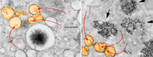
I have not really found the equivalent of the electron dense particles in the RER of Gclc mice (posted yesterday) for conditional liver ko animals rescued with NAC at 60d. I have also not found an equivalent (googling and other pathology type searches) for the iron spicules found within those partially RER vesicles.   However I did come close to finding a match for the vesicles within vesicles (though these cells were in vitro, not liver, and not in vivo). The partial publication URL which shows iron particles (USPIO nanoparticles at iron concentrations of 50 μg/ml)(link here) which are NOT LIKE the spicules found in the mice I am looking at, but does show something like the vesicular changes, that is, the vesicle-within a vesicle configuration.

I have pseudocolored the vesicles to compare orange, and on the left is the image from the Gclc conditional ko rescued with NAC, and on the right is the cell line (Canine ADSCs or canine adipose derived stem cells) treated by these researchers with iron.  Red arrows point to the vesicles within vesicles, black arrow from their micrograph point to nanoparticles of iron.  Heavy metal (maybe iron in left hand micrograph) looks like a fuzzy black iron filing mass pulled by a magnet into a glump, very different from the iron in the micrograph on the right. It is critical to point out that the vesicles are pretty much ribosome free, while those RER bound objects with electron dense (presumptive iron) have ribosomes, at least over part of their surface.

electron microscopy liver possible iron deposits vesiclesHere is another example of metal (in this case aluminum) in tissue culture forming dense lysosomal bodies.

Electron micrograph of liver: Alb w/c Gclc i/i D60 NAC rescue – color

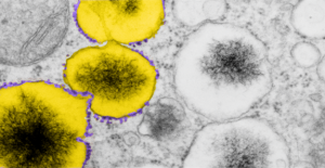
A portion of the previous post has been enlarged and also pseudocolored in photoshop. The ribosomes from this portion of an hepatocyte are colored purple, this is mainly to emphasize that whatever these vesicles are in the cytoplasm of the rescued mice do have some kind of protein being produced.  The portions of the vesicle membrane that are occupied by ribosomes is not that great, when found, too, they assume a position as if a string of 4-8.  The vesicles in tandem, themselves are colored bright yellow.  The contents of the vesicles needs no coloring it is very electron dense and likely to contain iron, be iron, or some other combinations of metals.

17902_74138_706_wcii_nac_60d_(colored).  Please note that on the right hand side, unretouched vesicles and inclusions are found, just in case you need to check. On the left hand side of the lowest vesicular blip, find three very nice ribosomes, but the remainder of the vesicle has but one or two additional ribosomes about the periphery.