Category Archives: Electron micrographs of liver

Electron micrograph of liver: Alb w/c Gclc i/i D60 NAC rescue

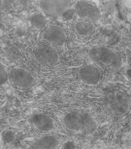
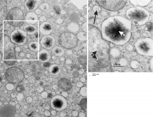
The four (maybe there are more) things that I noticed about the GCLC conditional KO mouse hepatocytes were 1: vesicles within vesicles, 2: lucent areas of mitochondiral matrix adjacent to quite well organized stacked cristi, 3: iron-like staining spicular deposits in dilated RER of hepatocytes, and 4: there is a linked-look between the ballooned areas of the ER (both smooth and rough).

These mice have survived the KO of GCLC, for 60 days, because they were rescued with N-acetyl-cysteine (NAC). NAC reportedly helps replenish the intracellular glutathione supply and can lessen hepatocyte oxidative damage occuring with the knock out of glutamate cysteine ligase catalytic subunit (Gclc), explained HERE. NAC affords protection against the oxidative stress, DNA damage and cancer development and cell death, thus rescues a genetically determined depletion of hepatocyte glutathione. I examined a large series of mouse tissues from these animals and their appropriate controls and while some of these data were published, there was much that never made it into the literature. Thus…. these posts, hoping that someone can be helped by these electron micrographic images of the unrescued, the controls and the NAC-saved mice.

So the micrographs below have RER (albeit the ribosomes are really not that nicely arranged in spirals or grids) and the transition from RER to ER is not consistent with typical RER to SER transitions (that is; both small and large vesicles, empty and full of electron dense filaments; those with center vesicles and those without (almost like continuous segmented mini-intussuceptions, are kind of a large mix of different types, sizes, filled and empty vesicles. These vesicles begin to develope before D60 after NAC rescue, and increase with increasing time.

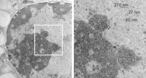
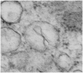
negative block and animal and treatment: 17902_74138_706_wcii_nac_60d. Size of a ribosome (at 27 nm) was used to calculate the bar markers (smallest black dot below. Inset from image on left is bounded with a white box, enlarge just around 2x on the right). Black arrow points to vesicle-within-a-vesicle phenomenon which is totally everywhere; double headed black arrow points to two types of mitochondrial inner membrane organization, one part points to areas which are quite electron-lucent, and one points to cristi that are highly layered and flattened. White arrow points to filamentous electron dense (i would like to say “iron filings” – like) inclusions and the grey arrow points to a telescoping point between two rounded contiguous vesicles with filamentous inclusions.  Occasional ribosomes are seen on the bounding membranes of these vesicles, and as in this case, they look like peristaltic connections, dipping one end into the next rounded vesicle as if there were strictures, or boluses passing through a long undulating series of tubes. Thee are a few coiled ribosomes on an mRNA through out the cytoplasm, and not pictured some microbodies (dense bodies with uricase crystals). Image to right shows the open-closed RER, SER, ER profiles show internally.
Fibril, filament, snRNPs, granular component etc

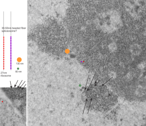
This micrograph I am using to determine the sizes of the different nuclear structures, so this is the first attempt to define at least four different sizes of components. For me, there is similarity in the size of the granules (beads on a string maybe something like 30 nm) in the cajal body and in the nucleolus. Arrows point to the kind of layered banding parallel strand look of the cajal bodies,  the red circle is about the size of a cytoplasmic ribosome (which would be something like 27nm) and the diameter of the rounded object beside the cajal body (something on the order of 60nm) and an even larger fibrillar component seen through out the fibrillar centers and also just a little above center left (orange spot) which might be 130 – 150nm.  So there are four measurements of fibrillar components in this nucleus, so far.  Will post more.

electron micrograph liver 14CoS ko nucleolar architecture

Fetal liver: electron micrograph

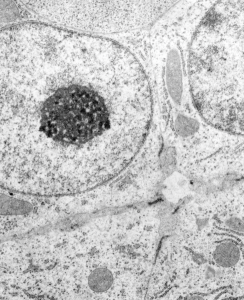
Fetal liver: electron micrograph which is pretty unremarkable. This particular image is take from a fetal rat at 20 days, but the mother had been exposed to dichloromethane during days 7- 20 of a timed pregnancy.  It has a great nucleolus for sure and the mitochondria are quite nicely7 preserved, the hepatocytes themselves don’t have a lot of stacked RER, and the bile canaliculus is really well organized and the little microvilli and junctional complexes among the four hepatlcytes pi9ctures are unremarkable. There is a cell at the top of this micrograph which looks to be an hematopoietic cell (probably part of an erythroblast). (57

While I did some electron microscopy for this study I don’t believe it was ever published. The PIs were J Manson and B Hardin, and some literature does exist on their experiments.

Nuclear pores: more than usual

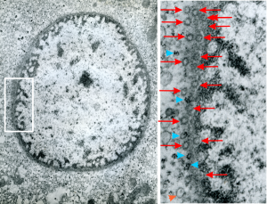
Hepatocyte from a 14CoS -/- mouse (homozygous for a 3800kb deletion on chromosome)that has spent 360 days being rescued from certain demise by NTBC has little condensed chromatin, very round and active looking nucleus and literally thousands of nuclear pores, more than I remember seeing in the nuclear envelope of any other cell type, and any other experimental condition.  Totally awesome. I would not hazard a guess as to how many are there per micron sq.

The purpose of nuclear pores is of course do provide a mechanism for selective transport of molecules in and out of the nucleus, separating the compartment of the cytoplasm and nucleus.  All produced proteins from the cytoplasm required by the nucleus will be imported through the nuclear pore, and the RNAs, mRNA, tRNA and rRNA, destined to become cytoplasmic machinery, transcribed and modified (post-transcriptional modification) in the nucleus will be exported to the cytoplasm through nuclear pores.

So the electron micrograph below (and inset) show this massive number of nuclear pores, particularly on one side that is cutting through the nuclear membrane at a slightly tangential angle, and also peculiarly, the polysomes (clear spirals of mRNA and rRNA) on the cytoplasmic side of the outer nuclear membrane are just about the only place in the cytoplasm where one finds RER. This vesicular ultrastructure (and particularly the vesicle – within – a vesicle) has been published Deiter et al 14cos, but not the nuclear pore observation. Here is such an hepatocyte nucleus, and inset to show tangentially cut nuclear pores (arrows), and spirals of mRNA with ribosomes (arrowheads). 17709_65053_14CoS-/- 360d w NTBC.

14CoS liver electron microscopy nucleus nuclear pores rescue NTBC


rRNA transcription just confusing

This is so awesome, just read part of this article by cheutin et al_awesome nucleolar diagram and view the TEMs and diagrams, were that I were beginning a career in TEM tomography, i would be in heaven. But 40 years too early.  So at best, is to take the new information and correlate it with studies from the past, making use of the data that I have and thousands of electron micrographs to play with.

TO begin with, 14CoS nuclei and nucleoli with some bar markers, just getting acquainted with the possibilities for labeling these structures. So here is the image from a previous post, and the segment enlarged with the two cajal bodies, tiny red circle is the size of a ribosome. Nucleolar structures I have not measured yet, but according to the publication by Cheutin et al, linked above, these will be similar and important in the pre-apoptotic hepatocyte nuclei found in this mouse line when they are not rescued by administration of oral 2-(2-nitro-4-trifluoromethyl-benzyol)-1,3-cyclohexanedione (NTBC).

electron micrograph liver 14CoS ko


14CoS: liver cell, electron microscopy

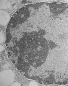
Really nice electron micrograph of liver nucleus and enormous nucleolus, lots of nuclear pores and perichromatin granules and condensed chromatin. 16029_65718_no_NTBC_14CoS/14CoS ko

Abstract from a paper by Deiter et al, follows: Whereas ch/ch wild-type mice and ch/14CoS heterozygotes are viable, 14CoS/14CoS mice homozygous for a 3800 kb deletion on chromosome 7 die during the first day postpartum. Death is caused by disruption of the fumarylacetoacetate hydrolase (Fah) gene; absence of FAH, final enzyme in the tyrosine catabolism pathway, leads to accumulation of reactive electrophilic intermediates. In this study, we kept 14CoS/14CoS mice alive for 60 d with oral 2-(2-nitro-4-trifluoromethyl-benzyol)-1,3-cyclohexanedione (NTBC), an inhibitor of p-hydroxyphenylpyruvate dioxygenase, second enzyme in the tyrosine catabolic pathway. The 70% of NTBC-treated 14CoS/14CoS mice that survived 60 d showed poor growth and developed corneal opacities, compared with ch/14CoS littermates; NTBC-rescued Fah(-/-) knockout mice did not show growth retardation or ocular toxicity. NTBC-rescued 14CoS/14CoS mice also exhibited a striking oxidative stress response in liver and kidney, as measured by lower GSH levels and mRNA induction of four genes: glutamate cysteine ligase catalytic (Gclc) and modifier (Gclm) subunits, NAD(P)H:quinone oxidoreductase (Nqo1), and heme oxygenase-1 (Hmox1). Withdrawal of NTBC for 24-48 h from rescued adult 14CoS/14CoS mice resulted in severe apoptosis of the liver, detected histologically and by cytochrome c release from the mitochondria, increased caspase 3-like activity, and further decreases in GSH content. In kidney, proximal tubular epithelial cells were abnormal. Human hereditary tyrosinemia type I (HT1), caused by mutations in the FAH gene, is an autosomal recessive disorder in which the patient usually dies of liver fibrosis and cirrhosis during early childhood; NTBC treatment is known to prolong HT1 children’s lives-although liver fibrosis, cirrhosis, hepatocarcinoma, and corneal opacities sometimes occur. The mouse data in the present study are consistent with the possibility that endogenous oxidative stress-induced apoptosis may be the underlying cause of liver pathology seen in NTBC-treated HT1 patients.

Electron micrograph: liver: Alb w/w Gclc i/i D120 NAC

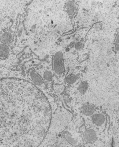
Electron micrograph: liver: Alb w/w Gclc i/i D120 NAC. So here is a perfect example of not having the right data written down while doing collaborative ultrastructure on a project for a colleague.  So after so many years I have to assume that this is a control liver receiving NAC for 120 days after birth, for a conditional Gclc ko mouse.

Liver looks really nice, WNL in my opinion, nice RER, nice mitochondira, one part of a lipid droplet visible. I partially hid two pen marks on this micrograph (using the bandaid tool in photoshop) which you can still see, but this did not change any data. Contrast enhanced as well to even up the tone of the liver histology.  liver, neg 17909, block 74119, anm#702, alb w/w Gclc i/i NAC 120 days post partum. 10000x orig mag. No profile of nucleus in this image.


Mitochondrial cristi: what is this ordered protein

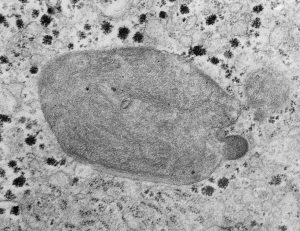
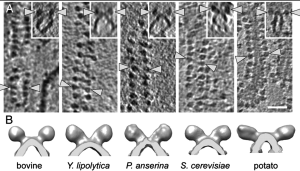
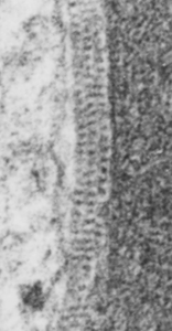
Mitochondria from vinyl chloride exposed animals have been posted on this blog before. HERE and HERE. I have made the assumption (no proof what-so-ever) that this just seems a perfect match for ATPsynthase.  Here is another example of such a mitochondrion, also showing the highly organized repeating pattern found in the intra-cristal-space of mitochondria from other liver electron micrographs from guinea pigs treated with vinyl chloride.  Amazing similarity. THis waits for someone else to confirm, but in the meantime when I see the images, I will post. Someone out there will know.  So here is a mitochondrion with such a cristi, and below that is someone elses micrograph (publication site linked) which shows the spiral repeating structure of ATPsynthase.


REF
One of their figures (not pictured here) shows about 16 dimers per 200 nm = 1 per 12.5 nm. When I measured the ribosome at 27 nm in my image (to the left) of intra-cristi structures, then there were about 19 densities per 200 nm.  So the numbers don’t add up too well.  Particularly with one of their figures copied above where their measurement (white bar in the figures and insets above) is noted in their publication to be 50 nm. This is quite far off what I see in the intra-cristi space in the figure to the left. It also seems clear that they have a propensity to be found in these groups between the outer and inner mitochondrial membranes and less frequently on the inner cristi.

Cajal bodies: associated with high levels of transcriptional activity

Cajal bodies (that have many other names as well, best one might be nucleolar accessory body) typically have a coiled architecture. I wonder if this is part of a necessary architecture, or whether is has been recognized as such because it was easy to identify, but Cajal bodies could possibly exist without such coiling, or better yet, maybe there are several different steps in the coiling in Cajal bodies, a a coarse coiling (50 nm thread width) and a fine coiling (5 nm thickness). I vote for the latter.  I think the rounded shape and polar position on the nucleolus, and the whole Cajal body size (it is a pretty large nuclear structure and was discovered first by light microscopy – the resolution of a structure there would be about 0.2 microns… so 200 nm).

My points: logs of transcriptional activity is found in neurons, in dividing cells, in cancer cells, but it has been my observation that there is also lots of activity in apoptosis.  It is something I hope to pursue, that is, whether the rounded structures, perinucleolar, the right size and shape for a Cajal body, but with out the coarse thread-look) might be a variation on a Cajal body, in anticipation of taking the cell down the route to apoptosis.

Really frustrating search for other images of Cajal bodies online, and one article published in a respectable journal talks about Cajal bodies WITHOUT an ultrastructural pix — who does that.

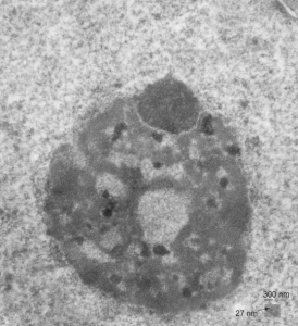
So this nucleolus is in an hepatocyte, the hepatocyte is highly active, there is little condensed chromatin the cytoplasm is full of vesicles, and mitochondria show oxidative stress, all in all a pretty taxes cell, and the reason is that it is a Gclc ko mouse, at 28 days, without any rescue antioxidant treatment and the hepatocytes are in the process of undergoing apoptosis. So this particular nucleolus, shows no dense fibrillar component around the fibrillar centers, and it has dense areas NOT the dense granular component, ut much more coiled and dense, and i think they look like Cajal bodies (but these would be bodies that do not have the 50 nm coiling, but seemingly the very much thinner filaments, those of 5 nm)  this is speculation at the moment, but I will add more info.

17740_73229_#203_mouse_wc/ii 28d, no NAC.   The dark round thing i think is the Cajal body is many hundred nm  in diameter,  bar = 300 nm, based on the size of several ribosomes (estimated at 27 nm diameter)(inset).

nucleolus_gclc_ko 17740_73229_27d_noNAC

Nice electron micrograph of a nucleus, nucleolus: mouse hepatocyte

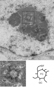
17531_73299_203_wcii_28d_nucleusThe nucleus is a terrific place to do electron microscopy. Aside from the non-membrane bound elements within which increase the difficulty for any attempts at reconstruction in 3D (at least in my mind) the micro-environmental pressures push this dynamic organelle to rapidly change and adapt. The nucleus here had one particular area that caught my eye, that is the fibrillar center – dense fibrillar component (within the black box) of the top figure. The electron micrograph is from liver and it shows a fibrillar center with a circular (perhaps more octagonal) shape (as do most fibrillar centers) but the arms of the dense fibrillar component extend in a rotational pattern. In this case there are four long spiral arms and four short spiral arms (think “galaxy” with spiral arms). This is not seen often and it may be chance but not likely, in my opinion, particularly when one views the old time lapse videos of cells in culture and sees the nucleus and nucleolus spinning like little worlds in the medium called space. Rotational forces are alive and well within the cell and it is not a big stretch to witness their effects.

That said, here is a little fantasy (all else preceding is for real) — would galaxy arms of the dense fibrillar component in all cells go the same direction?, would it be clockwise, would the dense fibrillar component drag off to the counter counter-clockwise south of the equator,  ha ha. For real, this is not a pattern I have seen described in the literature, nor recognized in the thousands of electron micrographs of my own, and thousands of others viewed, but I predict it will appear again and again.

Top figure is the original micrograph of a mouse hepatocyte,  exposure evened in photoshop, one tiny scratch is patched (dare you to find it) without affecting interpretation. Black box surrounds the nucleolar fibrillar center and spiral arms of the dense fibrillar components which were copied and enlarged from the original micrograph.  Inset lower left: burn tool in photoshop enhances direction of the galactic arms of the dense fibrillar component (which is visible in the original before emphasis), light central area is the fibrillar center.  Diagram in lower right – stick figure for orientation, FC, fibrillar center of the inset to left, DF dense fibrillar component (galactic arms), and GC granular component. 12531_73229_#203 wc/ii liver 28d.