Still going through all the transmission electron micrographs of alveolar type II cells from 40 years of photography. Here is one print from an owl monkey (no treatment) I pseudocolored with yellow-brown for the nucleus, red for the nuclear pores, blue for the lamellar bodies, cyan (green) for the cytoplasm. Just for the record, i did not find any protein structures within the RER which might be construed as intrcisternal layered RER inclusions that one wants to see if one is looking for SP-A.
Monthly Archives: June 2016
Owl monkey type II alveolar pneumocyte
I have searched, with electron microscopy, several different mammalian species for a pattern in the RER of protein which might be considered an accumulation – paracrystalline inclusion like thing comparable to what I have seen in guinea pig, ferret and dog, but none seen so far in owl monkey. Here is a cut and pseudocolored image of a moderately good type II cell from an owl monkey…. taken for another study (anm #1 neg 7160 bl 24060, untreated routine TEM) in 1982. Lamellar bodies are mostly empty in this particular image.
The nucleus is pretty dense, lots of condensed chromatin on the right hand side, and perhaps a portion of the nucleolus is there. There are some dense blue granules (presumed to be glycogen) near the bottom of the type II cell and also scattered elsewhere. (The type II cell was cut out using the eraser tool and lasso tool, and dirt and scratches were removed with the “bandaid” tool in photoshop).
5 year old mongrel dog lung endothelial or alveolar type II epithelial cell? inclusion
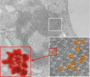
From the previous post, I did some more searching on the very ordered protein inclusion in the type Ii cell of a mongrel dog (5 yr old). The orderly array looked to me like a bunch of hexagonal structures with a central dot. When I made measurements (comparing the magnification, enlargement and pole piece used at the microscope, I figured out what about 500 nm would be, and then 50 nm seemed to be almost exactly twice as big as each of these hexagonal flower (bouquet) structures. They might not actually be octadecamers (as is often found for SP-A )- and a shadowed image of one such shadowed SP-A molecule from a publication by T. Voss, H. Eistetter, KP Schafer in J. Mol. Biol, 1988, 201: 219-227, which i have cut out, pseudocolored in photoshop, cut out, and placed along side one of the bouquets in the ordered protein inclusion in this particular cell. Since I could not identify the cell specifically, one only can guess as to what it might be.
nb. since this blog, I found a similar micrograph of dog lung, in a paper by Stephens, Freeman, Stara and Coffin, Am J Path, 73: 711-726, 1973 which identified a very similar structure (cuboidal organization) in endothelial cells (they say)(Figs 14-16 in their publication). I cannot identify the cell in my micrograph as an endothelial cell, but can confirm that there are dense coated vesicles (at least 11 in this image) just like they found in their micrograph., and one edge of the cell in my micrograph is adjacent to collagen fibers. This would make the cell either epithelial or endothelial. It has no lamellar bodies and no nuclear profile, and only two kind of iff-ish mitochondria.
Circles identify the two molecules (one SP-A according to Voss et al, the other a similar sized protein (could be an octadecamer?? just making a guess here) or some other order of –merization. In the two images (see previous post) there were literally hundreds of times that one could visualize a bouquet with center point as the one enlarged in the inset, and pseudocolored a purple. One of the perplexing things is that when one begins to see structures in a repeated design like this…. many more can be pointed out, and then one wonders what is imagination and what is fact (that is the great tendency of the human brain to become immediately biased).
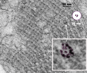 So after finding this in the publication by Stephens et al, 1973, I decided to scan another area of that same cell and enlarge a portion, and highlight some structures (as above) which look like a multimer of SP-A in a lattice formation and post it here again. Would love to say I could count 18 blobs to depict the carbohydrate recognition domains (haha). I highlighted the “spokes” found in the inclusion below, I should have seen them in the inset inclusion above, but did not highlight them… however, they are there.. maybe 6 of them in top, maybe 7 in bottom inset. The configuration here , globby tops, skinny necks and central dot is pretty reminiscent of how the bouquet of SP-A is diagrammed.
5 year old mongrel dog lung RER inclusion ???
These accumulations are found in the peripheral lung of a 5 year old dog that was part of research for an entirely different topic. The identification of the cell was not really possible, there were not absolute defining features, except there were mitochondria, perhaps a lipid droplet (cell membrane is obscure around this area), dense coated endocytic vesicles, rough endoplasmic reticulum, and it is adjacent to a cell(s) with microvilli, basement membrane, and alveolar space. What it is, I have no clue.
neg 9931, blcok 24669, dog 5 yr, original mag 21750 x 4 pole piece V seimens 1A
Early efforts at morphometry :)
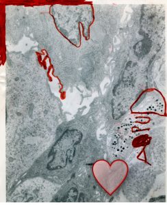
Long ago, my daughter was frequently at work, and here, as reminded of by finding this micrograph in my stash, as a young child she must have been visiting my office – maybe school was on a “teacher’s” day, or some other issue was at hand. Many of the methods for morphometry I have used (and developed) over the decades (to determine organelle sizes, incidence, etc) she must have been aware of. Here she correctly outlines the nucleus, the cell plasma lemma, and in particular she liked the granules in the lower right (ha ha) I do not remember what APUD stood for that day. This micrograph I must have given her to entertain her…and teach her that science can be fun, and also can be for women (believe me, when I was in graduate school it was not a happy place for women), and it can be artistic. I don’t remember how old she was, but not very old. The heart is “mine” in finding this memory.
Microvillar cell in the olfactory epithelium and alveolar type II cell in lung: RER look-alike
Thinking about dendritic cells, in general, which participate in innate immunity as I have done recently because of my efforts to put a “name” on the protein responsible for the layered RER granules in some alveolar type II cells. I have searched the micrographs of many tissues which I have examined over 40 years, and the widespread similarities in morphology, one cell to the next – because of similar mechanisms for producing – importing – exporting – secreting – excreting -recycling – removing – transferring – deleting – various “products” as granules – proteins – factors – hormones – etc, are pretty interesting. In particular, such varied products come from such uniform mechanisms of production. Case in point — the microvillar cell of the olfactory epithelium (which I happen to think is a dendritic cell with functions related to innate immunity) has RER which in someways is a dead ringer for the RER of the alveolar type II cell in some of my guinea pig micrographs with RER granules that appear to be overproducing some protein — likely a surfactant protein, either A or D. See below…. Only by the presence of a lamellar body can these two cell types be distinguished… in particular RER profiles can often be amazingly similar (OR — alternatively are the protein products amazingly similar – maybe both are c type lectins). IF YOU FIND THE LAMELLAR BODY you will know which is which… LOL — I’ll never tell.
It is my hope that at some point the protein being secreted by the microvillar cell of the olfactory epithelium is a c-type lectin just like at some point the protein in my guinea pig type II cells will be determined to be surfactant protein A or D or both. I did hunt vigorously for any layering and periodicity in the former, but did not find any in my images, but found literally thousands of examples in the type II cells (see previous posts).
RER protein in a type II cell of a guinea pig
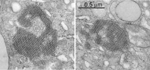
The stacks of RER cisternae in this particular type II cell don’t show the layering and patterning that other cisternae from these animals do, that is, they look more like ordinary RER cisternae (though clearly a little dilated, and clearly more stacked). Still I think this is an overproduction of SP-A or some SP as other images from the same animal show the periodicity seen in intracisternal bodies.
The stacks of RER are reminiscent of the stacks of birbeck granules in the Langerhans cells where langrin is overproduced. This is a paper by J. Valladeau et al, Immunity 12: 77-81 (2000) where they transfected langerin cDNA into fibroblasts and got stacks of birbeck granules (which they show in an electron micrograph). While the image below is certainly NOT langerin, and NOT a transfection of any SP-A cDNA into alveolar cells, it is probably the ONLY image that i have found where the profiles or RER might mimic granules (RER) of other c-type lectins (like the birbeck granule) in terms of being segmented and stacked.
Awesome polysomes in a guinea pig type II alveolar cell!
There is no underlying point for this, just was this wonderful coil of polysomes in a type Ii alveolar pneumocyte from a guinea pig and wanted to capture the “beauty”.
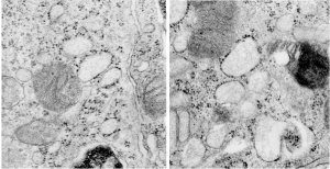
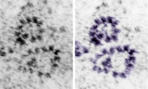
Original unedited micrograph (to estimate magnification, each ribosomes is about 20 nm in diameter) is grey scale (L) and a few of the ribosomes and the polysomes are pseudocolored purple (R).
Who has electron micrographs of c-type lectins in situ, in vivo
I am continuing to search for electron micrographs which have images in high resolution and magnification of the c-type lectins to compare with the intracisternal protein accumulation in type Ii cells of the lung of some species of mammal. Upon thinking about the, comparing with the fine structure of langrin in the Langerhans cell, it occurred to me that I might have a similar structure in the microvillar cell of the olfactory epithelium.
The microvillar cell has not been a topic of much research, and it has no well established function. Because of the occurrence of about 10% of most epithelia in the body to be burried in the epithelial cells proper (10% in olfactory epithelium, back skin, colon epithelium, etc) that I have examined personally it made sense to go over that set of about 250 micrographs looking specifically for RER profiles that specifically had a periodicity or layering to the protein content. Unfortunately, in all those microvillar cells examined, I did NOT find any organization to the protein contents of the RER profiles, but I did find the sparse ribosome studding on the profiles that I have seen in the type II cell of the lung.
Nothing conclusive, but just a little bit peculiar. I do wish I had seen some indication that the RER of the microvillar cell had a “granule” like the birbeck granule… ha ha…. that would have given an instant functionality to the microvillar cell. It might still be there… as not all RER protein contents show periodicity like the one in type II cells and Langerhans cells. I have not found images actually for any other collectin that speak to layering, just the one I have examined and the birbeck granule in Langerhans cells.
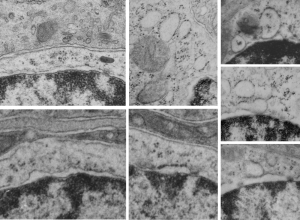
Here is a quick composite of some profiles of RER in the microvillar cell of the olfactory epithelium (recall, that upper layers contain the olfactory bipolar cells, the sustentacular cells (supporting) and microvillar cells (at about 10% occurrence. Profile of nucleus in the bottom sector of each image, perinuclear space and RER profiles are just above that. RER profiles are quite electron lucent, and only on e of the pictures shows any contents (and to me those are too close in shape to ribosomes to be excited about).
I would love to see any electron micrographs of MBL or other c-type lectin protein overexpressed in any type of cell.
Concentric tubular arrangement for surfactant protein in RER of type II cells
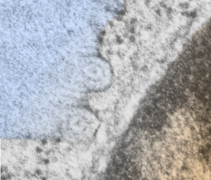
More images which pique the imagination about what is responsible for the highly organized protein in the RER of the type II alveolar cell in some mammals (in this case an aged guinea pig, control, from a previous study). I have mentioned before that there are examples of wildly converging and blending and mixing of these proteins going from a perpendicular arrangement to parallel to the long axis of the RER profile and splitting off and curving and also concentric or semilunar. This is a really nice resolution and relatively clear image of a tubular arrangement, cut in cross section, comprising a single “period” that is here seen as center dot, medium density ring and outer ring. This short video is constructed from two images, the initial image is untouched, the fade in image i have used photoshop “burn” tool to highlight the area that looks to me like it is concentric.
I added a blue color to an overlay of the actual cisternal body on the left, and a brownish overlay to the nucleus at the bottom right corner. Ribosomes are clearly found, and are especially prominent where the banding runs parallel to the RER, just above the tubular section.
Perinuclear space might have a trace of this protein in it, perpendicular to the direction of the nuclear membrane but it is quite indistinct, and there is a perichromatin granule very near the nuclear pore seen on the left hand side of this profile of the nucleus.
video is available on YouTube:
Composite image of intracisternal body (blue, nucleus, brown)


