there was a boisterous man, trump
who bombastically assumed he should stump
for the highest office the US possesses
he lied and he boasted and denied his excesses
with trumpistic ego and a trumpestuous grump
he trumped the opponent – no one would have guessed
it’s laughable and unsettling how his trumpastic mimes
feed media with countless hilarious lines
he fooled no one except those, the most trumpled
sadly I predict that their hopes will be crumbled
by their choice for a bully who lacks self-control
with an unbelievable ego and self-serving goal
he is unable to refrain from abusive attacks
on those who would challenge his ill-thought hacks
such as thinking its ok to avoid millions in tax
on his earnings through loopholes that should have been axed
whose trumpology doesn’t pass intellectual attacks
like immigration and outsourcing his plans have big cracks
those with a brain have long left his side
those who love humor hang on for the ride
elitists and intellectuals are allowing this man
to try to do what he thinks that he can
holding their breath with hope for achievement
and predicting slim odds they’re all in agreement
we will see if a trumpistic and hedonist mind
can bring about good changes if he takes time to find
an approach to the issues we have at hand
but not by trumping, grumping, and complaining
not with blame, belittling, and defaming
by avoiding excuses, from smoke screens refraining
a big order for a closed mind so small
I hope that he finds the strength to be tall
and gracious and kind and for seeking the best
for the america which I hope puts him to the test
america is great, I hope trump heeds the call
we’re not here for his ego – it is “america for all”
Monthly Archives: January 2017
Website front pages that are dominated by links not content
I am posting this rant, yes it is just that, an opinion, but I wonder how many share this thought. I really am annoyed when Kroger pops up an add on a fake stained glass pattern website that has nothing but links to other products and link backs to additional supportive webpages to trick google. I find it deceitful. I wont run google adds, even if i have to go out of business. They are ugly, intrusive, and cater to a clientele that does not have the viewers “experience” in mind. I do try to keep my websites clean, easy to navigate, and tell you exactly what you will get when you make a purchase. Please understand that for me, anyone who runs a stained glass website, ought by default, just to be a little concerned with their credibility as an artist…. Look below… I gladly hold my three sites up against any of the others listed. regarding appearance and ease of use…. it may not get me much in terms of click-through dollars (in fact NILL, ZILCH, NADA, OUGHT, ZERO) but it is not google-add-ugly or ludicrously link heavy Mine are the top three.


Cajal bodies: associated with high levels of transcriptional activity
Cajal bodies (that have many other names as well, best one might be nucleolar accessory body) typically have a coiled architecture. I wonder if this is part of a necessary architecture, or whether is has been recognized as such because it was easy to identify, but Cajal bodies could possibly exist without such coiling, or better yet, maybe there are several different steps in the coiling in Cajal bodies, a a coarse coiling (50 nm thread width) and a fine coiling (5 nm thickness). I vote for the latter. I think the rounded shape and polar position on the nucleolus, and the whole Cajal body size (it is a pretty large nuclear structure and was discovered first by light microscopy – the resolution of a structure there would be about 0.2 microns… so 200 nm).
My points: logs of transcriptional activity is found in neurons, in dividing cells, in cancer cells, but it has been my observation that there is also lots of activity in apoptosis. It is something I hope to pursue, that is, whether the rounded structures, perinucleolar, the right size and shape for a Cajal body, but with out the coarse thread-look) might be a variation on a Cajal body, in anticipation of taking the cell down the route to apoptosis.
Really frustrating search for other images of Cajal bodies online, and one article published in a respectable journal talks about Cajal bodies WITHOUT an ultrastructural pix — who does that.
So this nucleolus is in an hepatocyte, the hepatocyte is highly active, there is little condensed chromatin the cytoplasm is full of vesicles, and mitochondria show oxidative stress, all in all a pretty taxes cell, and the reason is that it is a Gclc ko mouse, at 28 days, without any rescue antioxidant treatment and the hepatocytes are in the process of undergoing apoptosis. So this particular nucleolus, shows no dense fibrillar component around the fibrillar centers, and it has dense areas NOT the dense granular component, ut much more coiled and dense, and i think they look like Cajal bodies (but these would be bodies that do not have the 50 nm coiling, but seemingly the very much thinner filaments, those of 5 nm) this is speculation at the moment, but I will add more info.
17740_73229_#203_mouse_wc/ii 28d, no NAC. The dark round thing i think is the Cajal body is many hundred nm in diameter, bar = 300 nm, based on the size of several ribosomes (estimated at 27 nm diameter)(inset).
Blurring the lines between organelles: RER and mitochondria tangential section
This is a great view depicting how closely aligned the outer mitochondrial membrane and the RER profiles can be. Smeared along a tangential section, opportunely showing about 7 ribosomes per spiral, (arrows 5) and also showing how close the RER is to the outer mitochondiral membrane and some of the cristi. So close, in fact, that it is not possible to define the separation between the two.
This sample is from mouse liver: indented (arrow 4) mitochondrion is indicative of oxidative stress, the RER components are dilated, some protein is seen, there are tiny vesicles within the RER profiles, ribosomes are not continuous (arrows 1 and 2), and have been lost at sites around each of the RER profiels. There are numerous polysomes, in addition to the ribosomal swirls attached to RER membranes. The increase in vesicles is a marker for the genetic disposition of this particular transgenic mouse. Mitochondria have a mix of tubular and flattened cristi, not that many intramitochondrial granules (Ca++ and Mg++). Right panel is an enlargement of black box in left panel.
Nice electron micrograph of a nucleus, nucleolus: mouse hepatocyte
 The nucleus is a terrific place to do electron microscopy. Aside from the non-membrane bound elements within which increase the difficulty for any attempts at reconstruction in 3D (at least in my mind) the micro-environmental pressures push this dynamic organelle to rapidly change and adapt. The nucleus here had one particular area that caught my eye, that is the fibrillar center – dense fibrillar component (within the black box) of the top figure. The electron micrograph is from liver and it shows a fibrillar center with a circular (perhaps more octagonal) shape (as do most fibrillar centers) but the arms of the dense fibrillar component extend in a rotational pattern. In this case there are four long spiral arms and four short spiral arms (think “galaxy” with spiral arms). This is not seen often and it may be chance but not likely, in my opinion, particularly when one views the old time lapse videos of cells in culture and sees the nucleus and nucleolus spinning like little worlds in the medium called space. Rotational forces are alive and well within the cell and it is not a big stretch to witness their effects.
That said, here is a little fantasy (all else preceding is for real) — would galaxy arms of the dense fibrillar component in all cells go the same direction?, would it be clockwise, would the dense fibrillar component drag off to the counter counter-clockwise south of the equator, ha ha. For real, this is not a pattern I have seen described in the literature, nor recognized in the thousands of electron micrographs of my own, and thousands of others viewed, but I predict it will appear again and again.
Top figure is the original micrograph of a mouse hepatocyte, exposure evened in photoshop, one tiny scratch is patched (dare you to find it) without affecting interpretation. Black box surrounds the nucleolar fibrillar center and spiral arms of the dense fibrillar components which were copied and enlarged from the original micrograph. Inset lower left: burn tool in photoshop enhances direction of the galactic arms of the dense fibrillar component (which is visible in the original before emphasis), light central area is the fibrillar center. Diagram in lower right – stick figure for orientation, FC, fibrillar center of the inset to left, DF dense fibrillar component (galactic arms), and GC granular component. 12531_73229_#203 wc/ii liver 28d.
Rough endoplasmic reticulum, stress responses in hepatocytes
Verge of a Dream: Someday you may paint me
 Someday you may
paint me. Not
in a scramble
to remember,
because not everyone must
feel they will miss
another point or
appointment. Maybe
not a portrait, sitting
slickly attired. Not
everyone must
think that what they
wanted, they misconstrued.
Just a picture of a wrist
with a monogram on an
eighteen carat chain.
Since you would
rather transform
than trade away.
You’ll buff up this
image with a palette
that does not give
away any secrets.
RB 01 04 2017
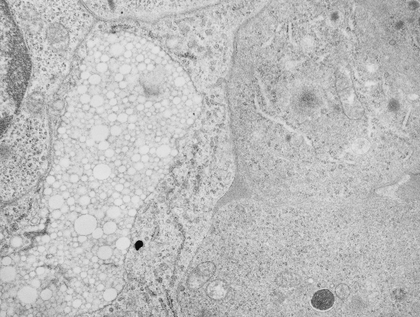
Perfluorotributylamine: re-emulsification within a lysosome
Electron micrograph of mouse bone marrow 100 days after the infusion of FC47 emulsion of perfluorochemical, in a study in the LC Clark, Jr, laboratory on artificial blood, blood substitutes. It has always been amazing that the original blood substitute-emulsion particles first are engulfed by phagocytes, and then demolished into seemingly amorphous inclusions, then with time, some perfluorocarbons, like FC47 are re-emulsified within the phagolysosomes into oodles (no clue how many millions) of small, very small and even smaller droplets. This particular electron micrograph of a bone marrow cell (just a portion of the cell) is packed with re-emulsified FC47 droplets within a membrane bound phagolysosome (for want of specifics on the enzymes in this organelle). It has been cropped and enlarged four times, and still there is no visible end of the increasingly tiny droplets. (Conjecture takes them to the level of the molecule. haha)
Arrow in top figure points to the small droplets that will be visible in the last of the enlargement inset figures.
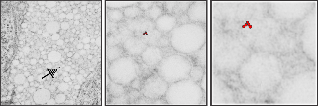
Mouse: emulsion FC47 = perfluorotributylamine number 800325EI, IP, 50cc/x 7 =350 cc/kg (one whopping dose), I would have to look up the mixture, but typically 10% by volume with 5%F68, neg 5402, block 16203, 100 days post infusion, bone marrow, orig mag 13900, V, x 4: transmission electron micrograph. To estimate size, if a mammalian ribosome is between 25-30 nm in diameter on the same micrograph, then the size of the smallest red perfluorotributylamine droplet in the last inset is something around 10 nm in diameter.
Perfluorotributylamine (FC47) emulsion: mouse, electron micrograph of bone marrow
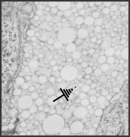
100 days after the invusion of artificial blood into mouse, this sample was taken of bone marrow. The white bubbly area on the lower left middle is typical of what perfluorochemical emulsions (at least those that eventually left the body) would look like inside phagolysosomes. The perfluorochemicals formed these small pockets in a sense re-emulsified within membrane bound structures in most cases (some perfluorochemicals like perfluoroiodide excepted). As a general rule of thumb, the faster the perfluorochemical blood went from large messy phagolysosome into a neat tidy highly ordered bubbly phagolysosome, the faster it left the body. I have been thinking on these images for 30 years, and hope to compare different chemicals in this respect. These are electron micrographic data from long past, before this type of blood substitute was approved and then disapproved for human use in the US. Nevertheless it tells us much about the immune system and processing of foreign materials (even man made materials).
 There are four types of instability in emulsions: flocculation, coalescence, creaming and Ostwald ripening…. all fun words that will likely apply to the transition of the first phagocytosed emulsion flocculant from the blood. I think in the case of the fluorocarbon emulsions there is a REVERSE Ostwald ripening in some, or alternatively, there is a minimum size that the lysosomal enzymes which are also within these phagolysosomal bodies can re-emulsify the fluorocarbon. (black dot is dirt… i didnt photoshop it out)
Guinea pig alveolar type II cell granule protein: surfactant protein A?
Guinea pig alveolar type II cell granule protein: surfactant protein A?
There are so many images which show the same data, layering, periodicities, hexagonal array at the growing ends of granules that the evidence that these are some regular sort of structure in select species is a little hard to ignore. I keep trying to convince myself that I am seeing what I think I am seeing. This opportune section of an alveolar type II cell (from ferret) granule is just kind of nice, as it shows the growing end ribosomes, the stiffness of the RER profile when the protein begins to organize (seen on the left hand side of the micrograph) the looseness at the growing end before further oligomerization takes place in the granule (right hand side of the micrograph) where it seems that there is some kind of organization soon after the protein leaves the ribosomes (like the hexagonal pattern for a top down view of surfactant protein A, as seen here (unretouched EXCEPT for the burn tool in photoshop to highlight what I have seen), though I would have liked to see more with the central dot. Ribosomes were burned to highlight, and a ribosome marker (nominal 27 nm) are given plus a bar marker at approximately 100 nm, and hexagons at the growing end of the granule are burned as well. Inset is enlarged from white box in top figure. 9835_17084_guinea_pig_#301
