doesn’t fit well.
doesn’t fit well.
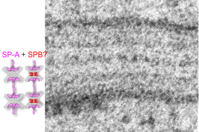
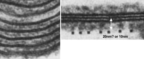
So do you think that SP-A is the main protein component of the alveolar type II cell granule? SP-B has been shown to work with SP-A in several circumstances and particularly in forming tubular myelin lattice structures. SP-B is a pretty small and flat molecule (but not as flat as DP-C, and it diagrams seem to place it wort of “surface-wise) to the plasmalemma bilayer. and if i remember right, maybe the bilayer elements of the alveolar space. Here is a cut and paste from a paper from the “before times” by Williams et al, 1991, where they show artifically created showing some lamellar ultrastructural similarities with the two dense lines and central punctate line seen in RER granules of alveolar type II cells. (but I am not sure about size) thought the mention of 20 nm is there, because i don’t know if that is a measurement from one dense lipid layer to the center punctate line, or all the way across the lamellae. Her reference is HERE. and a cropped figure from her publication is below. The match is certainly not perfect, but suggestive of possible structural organization of SP-A and lipid.
THERE IS STILL HUMOR!
LONDON (The Borowitz Report)—The theoretical physicist Stephen Hawking angered supporters of Donald J. Trump on Monday by responding to a question about the billionaire with a baffling array of long words.
Speaking to a television interviewer in London, Hawking called Trump “a demagogue who seems to appeal to the lowest common denominator,” a statement that many Trump supporters believed was intentionally designed to confuse them.
Moments after Hawking made the remark, Google reported a sharp increase in searches for the terms “demagogue,” “denominator,” and “Stephen Hawking.”
“For a so-called genius, this was an epic fail,” Trump’s campaign manager, Corey Lewandowski, said. “If Professor Hawking wants to do some damage, maybe he should try talking in English next time.”
Later in the day, Hawking attempted to clarify his remark about the presumptive Republican Presidential nominee, telling a reporter, “Trump bad man. Real bad man.”
I have tried to figure out how many square meters are involved in the interface of our environment in lungs and in intestines. I think it is fair given the low surface are of skin to consider it as secondary in m2 to the other two. I think i remember around 17 m2 or something close. But I have read 2 publications that have shown up in numerous lay publications as well that say the gut has less surface area than previously determined. The early estimates (that is, when looking at plicae, villi, AND micovilli – when they exist on epithelial cells in the gut) that the m2 was something on the order of about 7000. The similar number for lung was between 30 and 200 m2 . I don’t know which is right, but the numbers for lung appear now to be more around 70 m2 , and the numbers for gut are also around 200 m2 . this would make these wet surface areas equivalent…. I think that is hard to believe. I am not suggesting one can be had without the other (LOL), but the mere number of microvilli, on the epithelial cells in the gut just is staggering, and the microvilli on type II cells in the lung, just not the same (the gut microvilli are actually brush border type structures, rigid, numberous, close together) while only a few little plasmamembrane blips on the alveolar cells exists, and random floppy microvilli on alveolar type II cells.
Just for me it would be hard to think of them being any near the same, and even then with the reduced value for gut, (down from the 7000 m2 ) still is a greater surface area than lung.
THUS, gut is the greater surface area interface with the environment: so eat wisely, you are eating your environment.
Who is on deck to decide the color of Kroger food packages? How silly is it to make food packaging for cheese, and other products, match the color of M&Ms… what gives… food isn’t bright plastic color blue, or green, or red. I don’t know what kind of a marketing ploy this was, but it was ill-advised by a generation that doesn’t apparently know that the health of their own customers is at stake. This is like the tobacco companies…kill your customers. By this i mean, I don’t think nutritionists who ask us to “eat the rainbow” of foods mean to buy the same color cheese in rainbow colored packages. Where is morality of Kroger-Corporate whatever. Rainbow packages are not a substitute for rainbow vegetables, but a ploy to entice purchases for the wrong reasons. I find their new packaging that focuses on anything except the product within the package, really unappetizing.

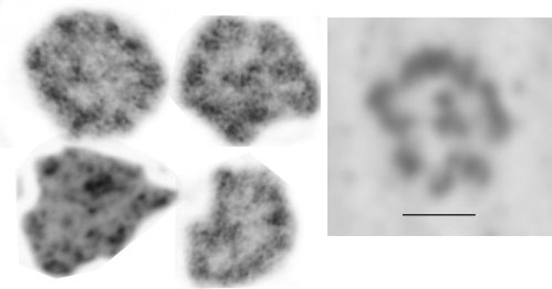
Voss et al, JMB 201 : 1988 published shadow cast images of SP-A (which I did a screen print on (right hand box with bar marker of 20nm for all images) and images from my own micrographs of the layered RER granule (intracisternal body) in alveolar type II cells found in some species. The bar markers are just a little different, which is not at all surprising, but the figures themselves and general size matches are pretty striking. I have compared with structures derived from tangential sections of RER granules (intracisternal bodies) in alveolar type II cells (4 samples on the left).
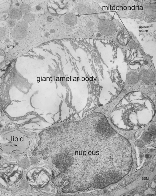
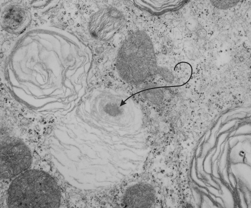 In lung, the type II cell has lamellar bodies, the secretion granule of surfactant, but not all species show a projection core within those lamellar bodies. I don’t know if guinea pig has ever been described as having projection cores in their lamellar bodies, but this looks either like a portion of a multivesicular body-fusion with lamellar body, or maybe a projection core (which should probably have more layered substructure). It has a different electron microscopic appearance than lipid does, and lipid droplets, seen as mostly textureless round areas, are found in lamellar bodies very frequently, and in many species in my experience. Here is a crop from a type II cell from guinea pig lung and the lamellar body (end of pointer) seems to me to have a projection granule. (6481_M8005 guinea pig #51, vinyl chloride exposed plus 2mg vitamin C you can access this pdf with specifics and details of animal exposures vinyl_chloride_vit_C_guinea_pig_lung )
I also went quickly through the electron micrographs I have for dog lung (467 lamellar bodies quickly counted, with 24 lamellar bodies equivocal for a projection core, but not really a good match, looking much more like areas where there is multivesicular body material.
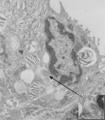
Also, perusing over 1000 lamellar bodies in electron micrographs from ferret my conclusion is that there are many little lipid droplets within lamellar bodies, but only a rare protein-like body that might under ideal fixation circumstances be seen as a projection body but still without substructure that is layered (which Ochs 2009 mentions is the hallmark for projection cores. He also states that they do not appear in rodents). There are many many examples of multivesicular type densities within lamellar bodies though, clearly defined and usually around the periphery of the lamellar bodies. That said, I understand that “fixation” is everything.
Here is an electron micrograph of ferret alveolar type II cell which has possibly as good an indication from archived photos as any that maybe in some ideal conditions, ferret might actually have some lamellar bodies with some type of density, maybe part of a multivesicular body-core together. I am not banking on it, but offer this, as one in a thousand example of a lamellar body that might have a projection core. As with guinea pig and dog, most of the roundish things at one or more poles or periphery of a lamellar body look more like lipid than layered protein. Lipid is common. This could just as easily be a tangential section of the bottom side of a multivesicular body fusing with a lamellar body. And only specific studies will determine which is which, and most likely will turn out to be something of both together.
I am posting this not really as a huge project, but just as a reminder to self, and to those interested that sometimes data are lost and forgotten but can still have meaning. The study mentioned here was just part of a very large study which was conducted by a good friend (late, actually many years passed on) who was very careful in what she did, and didn’t press conclusions or publications beyond their real findings. She was also a good friend. Dr. Martha Radike (who did these inhalation experiments at the University of Cincinnati, College of Medicine, Department of Environmental Health, back in the 1980s. The test animals (160 male Hartley guinea pigs) represented significant effort. I did some of the electron microscopy but never completed a manuscript on the animals (and they represented only 18 out of the sum total of the experiment.
My interest in the guinea pigs was revitalized while doing a bigger review of over 1400 micrographs looking specifically for a granule of surfactant protein in alveolar type II cells. So this study I brought up out of the archives for that purpose and found a summary, which I have posted HERE as a pdf for anyone who is interested. vinyl_chloride_vit_C_guinea_pig_lung
Here is an enlarged image from that pdf.