Monthly Archives: December 2016

Alveolar cell granule: perpendicular layers

Electron micrograph of a guinea pig alveolar type II cell which shows a granule of layered protein in the RER, which has a concentric arrangement (almost like a braided rug – for those of you who know what these are).  Along the periphery of the granule however, there are at least two areas, maybe three, where the layering (banding) has branched out perpendicularly to the concentric or wrapped original granule.  black lines indicate the direction of new layering nidi, and the scalloped lin shows areas which are lilely to be the marking points of each “period” bar marker is 270 nm based on the dimensions of a ribosome from this same micrograph.  96813_17084_guinea_pig_301 (untreated).

9813_17084_gpig_301_ICBa

Inner nuclear membrane: rigid and flexible portions

Nuclear membrane: rigid and flexible portions. I have noticed for some time that there are differences in the “lay” of the nuclear membrane (that is the inner nuclear membrane). There are stretches of inner nuclear membrane that are rigid looking, generally are devoid of condensed chromatin, still have the lamins present, but the protein groups attached or adjacent to the nuclear lamina of the inner nuclear membrane are spaced at a greater distance than the proteins in places of chromatin condensed along the inner nuclear membrane.  I found this particularly nice area in a guinea pig alveolar type II cell nucleus (which however had been exposed to vinyl chloride for a year), but this is a phenomenon not related to exposure but normal nuclear architecture. Cant wait to do more on this.

Below is an electron micrograph of an alveolar type II cell of a guinea pig, exposed as mentioned above to vinyl chloride, but this is just an image showing the inner nuclear membrane configuration which probably will end up being some kind of annulus around a nuclear pore complex, maybe part of the landscape that prevents condensation of chromatin in the nuclear pore area.

6476_M8005_gp_typeII_nuc_memb_stiff

Quick comparison: vinyl chloride exposure vs toluene diisocyanate in lung

Quick comparison: vinyl chloride exposure vs toluene diisocyanate in lung — alveolar type II cells in guinea pig. Not going into much detail, but just an observation after comparing two strains of guinea pig (Hartley and English short hair (males)) from two different experimental protocols, each having their own age matched controls (from experiments performed way back in the 1980s by Dr. Martha Radike and Dr. Stuart Brooks (respectively) and looking just quickly at hundreds of images of type II cells (looking in fact for surfactant protein granules – an entirely different study) I would say that vinyl chloride exposure did two things quite differently than toluene diisocyanate. I am not going to dig up the exposure protocols, days exposure and stuff because it is published somewhere, but vinyl chloride definitely caused an increase in the lamellar body size, and a concommitant decrease in the amount of RER and other organelles in the cytoplasm. Lamellar bodies got very large, caused indentations to nuclei and sometimes fused into these really large lumpy lamellar bodies. In addition mitochondria got large, had cristae deformed by some kind of matrix issue, the electron density was not decreased in the matrix, but cristae looked pushed and squeezed, in addition to an occasional fluting of the outer mitochondria membrane. (BTW, liver mitochondria from vinyl chloride exposed animals did show differences from unexposed as well). Figure from neg 6476, block M8005, anm# 51, vinyl chloride, low vitamin C. See below, two great nucleoli, several mitochondria with diminished cristae to mitochondrial ratio, enlarged matrix, and odd shapes within mitochondria.  The RER in the cytoplasm doesnt fig the granular layered profiles that have been seen in guinea pig that I am trying to establish as surfactant protein A, but are dilated and atypical for an alveolar type II cell from an untreated, and well, guinea pig.

6476_M8005_guinea_pig_typeII_VC_lowVitC

Toluene diisocyanate on the other hand (and some of the control animals along with… so you can judge whether this is an animal room sickness affecting all guinea pigs or something unknown) did NOT show mitochondrial or lamellar body size increase.

Intra-cristae – membrane protein organized in mitochondria

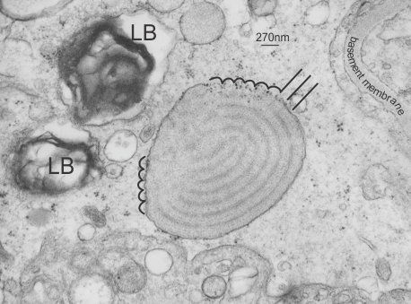
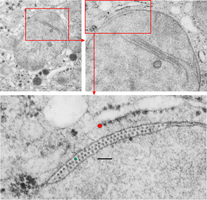
Intracristae – membrane protein organized in mitochondria — found in guinea pig liver electron micrographs in a retrospective study on vinyl chloride (and air) and vitamin C exposure from the 1980s. These cristae with apparent organizations of ATP synthase in a long row, parallel and sometimes looking staggered, sometimes aligned, have appeared in exposed as well as control (both groups receiving vitamin C). This is an awesome image with a 3-layered row of highly organized molecules looking a little like they are strung together somehow, yet each distinct. Just marvel at the order in the completely UNRETOUCHED electron micrograph  of a mitochondrion in guinea pig liver here.

4158_M8011_guinea_pig_liver_VC_vitC_HR
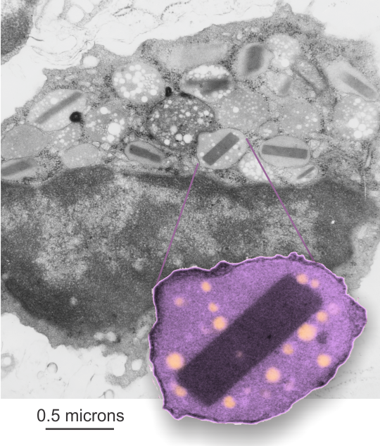
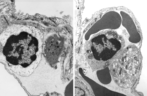
Mitochondria in both control and experimental data sets have mitochondria with wild shapes, very atypical for mitochondria in general, in some cases on section looking like asterisks (yep, same type outer membrane configuration seen in apoptotic cells). This is the case of the mitochondrion shown enlarged (the scalloped edges are just cropped so the ATP synthase is more visible. Image below 5924_17010_guinea_pig_air_vitC_liver.

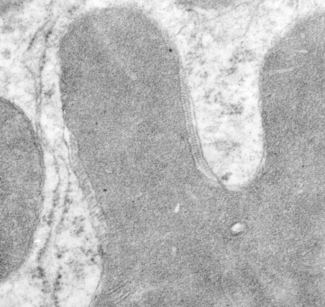
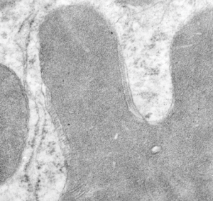
As with the intracisternal body in the alveolar type II cell of the lung of many guinea pigs, when the protein within the cisternae of the RER begins to assume a substructure (in that case, a layered granule), then the resulting RER becomes very rigid looking.  Similarly here, when the cristae have the ATP synthase all aligned in alternating double rows, the intramitochondrial membrane and other elements in the mitochondria also take on a rigid appearance.

5924_17010_guinea_pig_air_vitC_liver
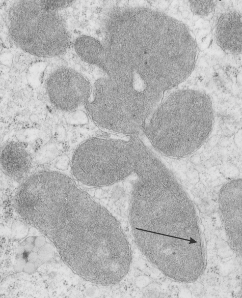
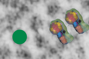
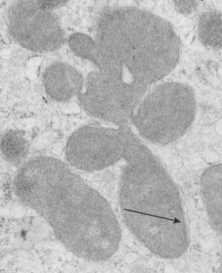
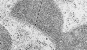
Micrograph below shows a section of an example of odd shaped mitochondrion with an area of extended ATP synthase molecules head to head, and on the inner mitochondrial membrane a little bit of rigidity and a thickening of the membrane is seen (arrow).

3548_M7991_guinea_pig_air_vitC_liver
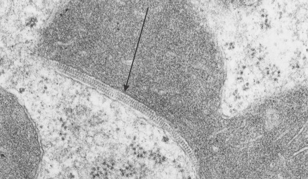
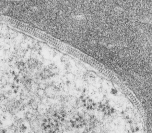
Here is a mitochondrion with an extended section of rigidity in the mitochondria membrane adjacent to a TRIPLE row of ATP synthase molecules, lucky cut (I hope that is what this is), and again rigidity in the mitochondrial membranes in this zone, and inner mitochondrial membrane has a slightly increased electron density about it (which of course means something, but I don’t know what).

4158_M8011_guinea_pig_liver_VC_vitC
HERE is a nice article…. it says the protein is in the matrix, which is not what is seen in the micrographs above, it is in cristae…. so there is some explaining to do on this topic.

Bias in long term memory.

When a president elect says something like “I won in a landslide” when he lost the popular vote and there were states that were too close for analysts to call the outcome for, then there is something amiss.

One has to consider the facts: wikipedia says (and wikipedia is a voice from millions of independent minds, perhaps the greatest collection of information ever created, free, and editable to all) — Memory gaps and errors refer to the incorrect recall, or complete loss, of information in the memory system for a specific detail and/or event. Memory errors may include remembering events that never occurred, or remembering them differently from the way they actually happened.

Wikipedia index goes on to list 13 different types of bias in memory, making it abundantly clear that we all fit into some of those categories, many quite deeply, others just barely. BUT not only is it intellectually dishonest to even begin to assume that one’s own memory is untainted, but it is also shows a total lack of understanding about biology and the mind, and how we store, retrieve and re-retrieve data.

What is just laughable (and also a serious problem for a country about to embark on a 4 year journey with a marginally fit mind at the helm) is that the comments, like the one above, fly out of a mouth with absolutely no frontal lobe editing. That is a scary thing.

landslide_victory_or_not

Dear MOM.

dear_mom    It is an odd thing to think about one’s life as it unfolds seemingly without effort ahead of one, but always requiring great effort to stay afloat.
I am constantly engaged in mental gymnastics trying to figure out the why and wherefore of it all and have not yet even come close to a satisfactory answer. It is hard to imagine another 25 years, possibly, ahead of me, not because it seems so long, but because it seems so short.
I have not learned how to reconcile my hopes and dreams with the reality of entropy and time, and I am like the cat with claws scraping down the wall trying to avoid hitting bottom.

Vinyl chloride and low Vit C exposure: guinea pig, liver, mitochondrion, intramembrane proteins

Vinyl chloride and low Vit C exposure: guinea pig, liver, mitochondrion, intramembrane, inclusions — a long title. This is an electron micrograph from the liver of guinea pig # 7 in a study about inhalation of vinyl chloride in conjunction with / without vitamin C. Dr. Martha Radike (who was a wonderful friend and colleague) did this study back in the early 1980s, but I don’t believe she ever published the results.  This particular animal (M8029) inhaled 600 ppm vinyl chloride 5 days a week for a year. The specifics of this which I cant remember (30 years ago) are 600 mmp VC, 4 hours a day, 5 days a week for a year, and this particular animal received 2-10 mg of vitamin C per day.  The mitochondria in lung, btw, were also very enlarged, though at this point I did not see any of these little inclusions within the mitochondrial membrane.

guinea_pig_liver_mitochondrion_vinyl_chloride_low_vit_CPublished reports have described changes from vinyl chloride exposure in liver: hypertrophy and hyperplasia of hepatocytes, activation and hyperplasia of sinusoidal lining cells, fibrosis of the portal tracts and the septa and intralobular perisinusoidal regions, sinusoidal dilation, and focal areas of hepatocellular degeneration. While the latter came out of a document that said DO NOT REFERENCE DO NOT QUOTE (oh well, excuse me), these are separate observations, but supportive of the unusual changes that Martha Radike (and I) found as mitochondrial changes seen in liver her year-long vinyl chloride exposure experiments in guinea pigs. There is a single mitochondrion pictured here, and two enlargements of areas (red boxes designate insets) which show the most wonderful inclusions I’ve seen twixt the outer mitochondrial membrane and inner mitochondrial membrane (I guess I could look for it in the intra-cristal membrane too but I haven’t yet). There are some misshapen mitochondria with cristae showing such in the air-breathing controls, which I will post, that received higher doses of vitamin C.  One wonders if the health of the entire colony played more into the results of all these changes (lung and liver both).

These little lollypops stuck on either side of the membrane are neatly alternating in a wonderful order and speak to something that I have not investigated but which was immediately apparent when I did googled “lollipops and inner mitochondria membrane” this may be ATP synthase. The dimensions are close to a reference I found: Structure of the mitochondrial ATP synthase by electron cryomicroscopy  by John L. Rubinstein, John E. Walker, and Richard Henderson.  There are some great videos on this molecular machine: HERE and HERE and HEREATP_synthaseIn the bottom section of the top micrographs the RED dot is @27 nm (ribosome), green dot @ 18 nm, bar marker, 100 nm. In the small bottom image i just cut and pasted two visuals of ATP synthase onto an enlarged portion of the above micrograph.  I added two charts to the pdf uploaded previously describing the materials and methods for vinyl chloride exposure. Find that document –> vinyl_chloride_vit_C_guinea_pig_lung1.

Next post is going to be a few pix of the ribosome bundle which is adjacent to some of these portions of intramitochondrial membrane with ATP synthase strings.

Mouse spleen: eosinophil after infusion of FC47

I am getting bored with surfactant protein A: Taking a break to look at some old micrographs from mice that were infused with FC47 (FC standing for fluorochemical) emulsions. This particular mouse (No. 8) was given 75cc/kg of an emulsion (10% in 5% F68 – emulsion number 741028) on four consecutive days (10-28-74 to 10-31-1974). The mouse weighed 22.2g  at sacrifice (11-1-1974), liver =1.79 g, spleen=0.62g. Tissues were fixed in Karnovskys, cacodylate buffer 2% OsO4, dehydrated in ethanols, embedded in Epon 812.

The unretouched image is grey scale in the background, one granule with perfluorochemical “droplet-footprints” remain in this (and many of the other) granules in this eosinophil. Components of the granules include histamines, peroxidase, ribonuclease (RNase), deoxyribonucleases (DNase), lipase, plasminogen, and major basic protein (stored in the dark crystalloid like structures within granules). Inset is contrast enhanced, cut out, enlarged, FC47 droplets pseudocolored.

The propensity for the minute emulsion particles phagocytosed by eosinophils to end up in the granules is something interesting in and of itself.  Several types of granules hold the little PFC (perfluorocarbon) footprints, not just those with MBP crystalloids.

mouse_eosinophil_spleen_artificial_blood_FC47


Mouse lung: liquid breathing E2

lymphocyte_plateletTwo archived electron micrographs from experiments in the 1970s which were part of liquid ventilation studies by Leland Clark, Jr.  These images are from a mouse that liquid breathed E2 (this is the hooker, I cannot find in my records what the chemical name for E2 is/was, but it is not what is currently searchable in google — what comes to mind is di fluoro methyl ether?).  This a mouse (typically swiss albino) was submerged in perfluorocarbon  for 15 min and tissues were taken at 0 min recovery.  Two micrographs of the dozen or so from this animal (I still have the Epon 812 blocks probably) and immediately I noticed two intravascular lymphocytes (probably medium size lymphocytes) making a quick pass by two platelets. One even has about 4 little surface folds reaching out to the lymphocyte.  I am not drawing any conclusions from two cells and two micrographs, but found it interesting, and would be highly surprised if something doesn’t point to this interaction, under some normal and experimental circumstances, as being significant.


Rat alveolar type II cell: just a nice electron micrograph

Rat alveolar type II cell, part of a nucleus , at least two nuclear pores (one a little tangentially sectiond, some good flattened cisternae of rough endoplasmic reticulum, lamellar bodies, polysomes, one coated vesicle (i think), some basolateral plasmalemma, collagen. One can see spacing of chromatin and or lamin proteins like little beads on a string… evne in the nuclear pore, perhaps two globular proteins right at the mouth of the pore ready to be polpped out into the cytoplasm.  See inset.

Rat, neg 9909, block 26538, 37500x enlargement 4 x.

rat_alveolar_type_II_cell_9909_26538