Monthly Archives: June 2017

Just a few tears for today

It sometimes overtakes me: Just the thought of the complexity of life as i see it through the electron microscope. An unbelievable maize of elements, most of which are still “unseen” and “unimagined” – one can spend a lifetime x 1000 building, searching, finding the patterns and puzzles of the ultrastructure of the cell.

Considering the tangle of filaments and fibers and fibrils and proteins as “beads” like pre-RNA, RNA, or nucleosomes or wrapped up like balls of string, fractal tangles (tangles is not the right word of course-as they are only tangles if one can not undo-them) and recursive patterns both physical and in DNA code, sub-structures that layer in unusual ways (SP-A, langerin, or anti-parallel helix loop helix proteins just naming three) that can look like little arms reaching out from highly ordered nuclear pores where it is possible even to see pre-RNA densities moving through the central portion of the nuclear pore, and in which one can imagine smaller molecules floating through on the outer-but still-inner confines of those 120nm wide windows.  Top down views of pores show rich octagonal symmetry (known by others to contain thousands of proteins), is extra special for the chromatin exclusion zone…. which provides little renaissance “arches” to protect, and to facilitate transport in and out of the nucleus.

And with hilarity, a mitochondrion can have as many (probably more) as three little dips of its organellar body, with outer nuclear membrane visibly pulld down to proximate nuclear pores to become exclusively entangled with the outer pore filaments.  And why is the nuclear basket on the inner nuclear membrane not that well defined in apoptosis? Is it that transport has become limited in cells preparing for self-destruct, closing up shop to preserve the well-being of the whole?

And this doesn’t speak yet to frequency of any of the very few selected items mentioned above as to mitochondria-cristi orientation and morphology, size and frequency; nuclear pores — frequency and morphology during high metabolic activity; and does not even begin to mention other organelles like golgi – size frequency, morphology, as perhaps, with the genetic absence of some magnesium transporter, or some sym or antiporters, channels, transport vesicles or influence of malfunction, or ER stress or environment… The whole thing is so complicated, so unknowable, so foreboding in its awesomeness and so frustrating to investigate, it just brings me to weep.
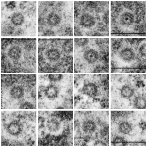
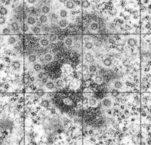
Nuclear pore gallery-1

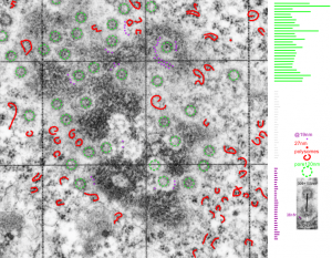
A collection of nuclear pores (top down) from a single tangential section of an hepatocyte nucleus from a mouse +/- for 14CoS (as opposed to the null). These show that about three quarters of the nuclear pores have some kind of transport item (like a pre-ribosome, or something else) being imported or exported all the time.  Just afun look at these adjacent pores. (excuse the morphometry grid from my inkjet printer back in the days when point counting was the method of choice for gathering numerical data from TEM images.

So these from a single cell show something about morphology… and the distance of the heterochromatin exclusion area surrounding the pore and the inter-pore distance and the size of the chromatin “beads on a string” in the adjacent chromatin I posted before.

Surrounding elements to nuclear pores: measurements

Some measurements, just made for my own information, might be useful in a relative sense to others looking at nucleus, nuclear architecture, and nucleolar architecture. Some data exist of course, but In the dataset that I am putting together from many different experimental protocols (archived) might lead to some new insights on numerical density of nuclear pores during times of cell stress, apoptosis, and in previously studies KO mice.  The numbers are meant to be “observations” and a suggestion.

This particular diagram has as its background  (top image) unretouched, and from a group of micrographs which were used for morphometry (hence the ink jet grid on the surface). It is hepatocyte, from a +/- 14CoS mouse which did receive NTBC (as a control for experimental mice receiving NTBC (data here) but showed pretty normal histology – but I predict with enough time and scrutiny something would show up. Octameric nuclear pores are shown as green dotted circles, ribosomes and polysomes presumably attached to the outer nuclear membrane, red, ribosomes, red = @27 nm diameter, peri-pore chromatin beads diameter, @19-20nm. Condensed chromatin at the inner nuclear membrane is well defined and “wound” in many places as a fibril also, possibly around 20+ nm diameter but longer lengths. About %66 of nuclear pores in this photo have central densities indicating some molecule is in transport one way or the other.

This particular animal had a great tangential section showing a large portion of the nucleus studded with nuclear pores and I used that opportunity to make some measurements on the diameter of the chromatin with beads (@19nm)-on-a-string appearance adjacent to the nuclear pores but outside the chromatin exclusion area around the pores and the distance between those beads (38nm); to measure the width of the chromatin exclusion area at places around the nuclear pores (maybe 2 -5 measurements perpendicular between pore and chromatin, per pore) (60nm+ 7nm and to measure the pore to pore distances as well (n=27, MEAN, 309nm+ 109nm (SD)).

Endstage apoptosis in hepatocytes

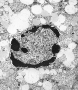
Here is a textureless (or nearly so) grouping of DNA around the periphery of an hepatocyte from a 14CoS ko hepatocyte, from a young mouse not rescued with NTBC.  Symmetry to clumps or peripheral digested DNA is obvious.  There remains some substructure to the “euchromatin” in the nucleus as well as some structure to the areas of presumptive digested chromatin.

Size and location of nucleolar bodies

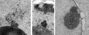
It is remarkable that these little segregated areas of nucleolar constituents look so consistently like “pop ups” that have a predetermined location, density, relative size, and texture.  I don’t believe that it is known what these early blips on the surface of the nucleolus are, (not including the blips that have the substructure notable of cajal bodies) though these may be precursors or similar structures. The banding (linear beads on a string appearance as if wrapped in a ball) in these bodies (by reason of tissue preparation or observer bias –shown in these three micrographs) would not qualify as cajal bodies. So what are they.  The bodies I am speaking of are all pointing upward like little volcanos (especially on the left)  with elements coming or going… whichever haha. Two images on right are slightly more electron dense than the adjacent nucleolar elements.

These images are from scattered micrographs, though two are from 14CoS hepatocytes which had never had or been withdrawn from NTBC.

Heterochromatin exclusion area around the nuclear pore

One cell from the liver of a control (sort of) for the Gclc ko mouse (that is that it had one wild type allele for the gene for turning on albumen synthesis (WC/ii  ) which rescues the would-be Gclc-/- mouse from death. This hepatocyte is not really remarkable to me but it did have this great tangential section of the hepatocyte nucleus and the nuclear pores were just scattered all over the area sectioned making the uniformity of the condensed chromatin areas surrounding the pores very visibly7 consistent.  It was so nicely consistent that I decided to try an afix a casual, but hopefully useful, measurement to that exclusion zone, at least in the this particular animal model.

So, that distance (based on the 27nm size of a mammalian ribosome turns out to be the following:  mean distance, 58nm.  From the original scanned image 58 nm+/- 16nm. That makes the space around the nuclear pore sort of a ring, of about 55 nm. Black line composit measurements, see bottomright. Red dot = one ribosome, electron microscopy nucleolus condensed chromatin exclusion

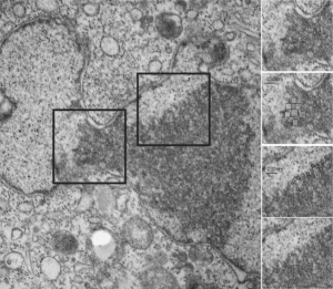
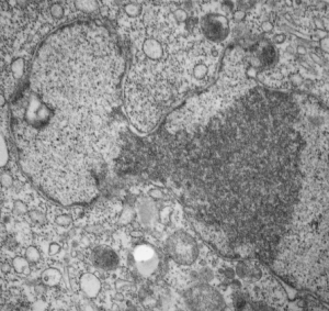
HeLa cell granular chromatin organization after irradiation with 30 joules UV?

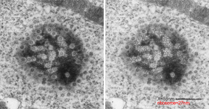
Here is kind of an interesting set of features in this clump of granular component of the nucleolus (or may just be condensed chromatin) that has a checkered appearance in some places (see checker boxes in inset micrographs to the right of the original image one showing the unretouched sections and an identical one with a checker pattern highlighted). There is a definite order to the spacing… or banding or filament pattern, something around 125 nm for the top two inset images, and for the bottom inset images, around 50 nm (which goes along with most measurements — being too big for interchromatin clusters granules (20-25nm), splicesosomes, around 30 nm, possible speckles? which are more like 50-100 nm).

HeLa cel electron microscopy nucleus

Nuclear pores: HeLa cells after irradiation with 30 joules UV

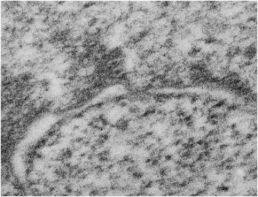
Nuclear pores: HeLa cells after irradiation with 30 joules UV — from an experiment trying to figure out how inhibitors of some of the caspases affected apoptosis. This study was from the 1980s and there are some interesting things never mentioned in that study which can be useful to someone out there doing electron microscopy on the nucleus.

There can be no doubt that some controls on nuclear size shape and activities are changed in HeLa cells… probably so different than the HeLa cells which started out way earlier than that. But while looking over “nuclear pores” in a different study I just happened to look over nuclear pores in this HeLa cell which had been exposed to 30j of UV.  I can find some evidence of nuclear pore cytoplasmic filaments but no nuclear pore baskets. So this is an N of 1, therefore not significant in and of itself, but when I googled nuclear pores and HeLa cells, there were publications, thus it is not an unusual, nor never-seen, entity.

Some of the central densities are missing from these nuclear pores as well.
nuclear pore electron microscopy HeLa cells irradiation Other interesting features include the “presumed” granular portion thee is a pattern, both on the upper margin before the “neck” (whose significance is not known) and also on the surface past the “neck”, I have put a checker board behind elements which appear to be organized into alternating space.  There are cytoplasmic filaments seen below but no nuclear side basket.  This is the most perpendicular section seen in this micrograph.

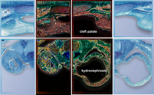
Cleft palate and hydronephrosis

Yet another set of images for a cover submission, this time for JBC but it was not accepted. I thought the microscopy with glowing edges was pretty nice, but apparently not to the liking of the cover editors.

The outside images look like they might have been plastic sections stained with toluidine blue (hence the little bit of metachromasia in the cartilage), the inside images are identical but photoshop was used to create the glowing edges.