This was a submission which actually did make it to the big screen. Gut and immunohistochmistry.

Monthly Archives: June 2017
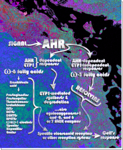
AHR. Probably was a cover submission, pseudocolored cell and diagram
Nuclear inclusion/invagination? which?
16040 65718 liver 14Cos 24 hr no NTBC, unidentified cell, not likely an hepatocyte, maybe an interstitial cell. Mitochondria in adjacent cells top left and right have condensed mitochondria. One of the interesting things about the “inclusion” in this nucleus is that it does not resemble any cytoplasmic structure in the cytoplasm of that cell, in fact the cytoplasm of this cell is quite different with lots of vacuoles (lower left) and an ill-defined shape, on one side very scant cytoplasm, which makes it less likely to be an hepatocyte than an interstitial type cell.
Three areas along the margin of the inclusion have been cut and enlarged (denoted by the three boxes in the original micrograph). The inset at the top shows there is really no distinct double membrane typical of the nucleus (inner and outer membranes and nuclear pores) and this might be a tangential cut-artifact. In the inset from the middle left, theer is a band of chromatin along the left border of the inset but again, no double membrane typical of nucleus. The inset from the lower right abuts up against nucleuplas which is very electron lucent, but along this area there is a density to the inclusion which looks like it might be some kind of lamin proteins.
Another great feature is the constriction appearing in lower middle of the inclusion, densities that look like they might have been some kind of “ring” around part of this structure.
Ribosomes (estimated at 27 nm in diameter from the cytoplasm have been enlarged with the insets (also shown in the original). Bar is 10x the ribosomal diameter at approximately 270nm.
The inclusion/invagination is full of robosomes and RER membranes (I guess that is what they would be named) with an homogeneous appearance and the vesicles or RER appear to be dilated to about the same uniform degree.
There is a nucleolus just above the inclusion. Animals with genotype undergo a pan-apoptosis among hepatocytes within a few days of birth, this doesn’t look like apoptosis yet, and also doesn’t appear to be an hepatocyte.
Perfluorophenanthrene in baboon lung
I don’t have a lot of data about the inclusions in this “apparent” alveolar macrophage (my best guess) but the round droplets seen at the left are perfluorochemical emulsion particles which is typical of what was seen in many species after intravenous infusion of such emulsions during studies on blood substitutes during the 1970s. This nucloeus caught my attention because I though perhaps I was going to find more “ring or donut” shaped nucleoli, but on closer look this area within the nucleus is infact a slightly curved invagination certainly caused by the pressure of one a smallish perfluorophenanthrene droplet on the side of the nucleus just below the section. Inset to right shows the nuclear pores which have been tantentially sectioned confirming that it is not nucleolus.
The range in size of perfluorocarbon droplets within cells is pretty amazing, and can range from sub-micron size to hundreds of microns in diameter. Size of particles plotted against time were related in some way to the off-gassing of the PFCs.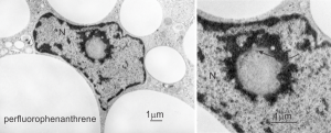 The widest medical use for perfluorochemicals today seems to be in ocular procedures for the detached retina (vitreous replacement?). Not for me. no thanks. Silicon oil would not make me any happier.
Chocolate donut and sprinkles: nucleolar ultrastructure
Shapes from biology (especially microscopy) sometimes are reminiscent of the most mundane objects in our daily lives. This nucleolus has reminded me of a donut with sprinkles for several decades. In searching the nucleolus for clues to the connection between the sizes of apparent granule-fiber, beads on a string, things to what has been reported in the molecular structures online, I used this particular nucleolus (from a baboon that had been given perfluorophenanthrene emulsion (probably IV)) and some tissues biopsied. This is lung. In other views, this particular cell (not clearly identifiable but likely an alveolar macrophage) contains PFC droplets.
The “sprinkles” for this donut represent the dense fibrillar component (entities measured on a post from yesterday; the cake part of the donut is represented by the circular structure in the center of the side-by-side identical micrographs, which is the granular component. NB, the granular component on the left is light grey and round with the fibrillar center being the center of the donut. It was noted in some of these nucleolar structures that there is such definite banding and that the lumps on a string is a very good casual description of the organization of RNP along a template and the measurements are pretty good. As a reference point the diameter of a ribosome is about27 nm and the space between ribosomes in this particular cell is around 58 (or 2x the diameter of a ribosome). The diameter of the beads in the granular portion is 21nm, with a distance separating them of from adjacent structures is quite large and prominent, around 42nm (twice the diameter of the granule and though smaller in overall size than the ribosome and its space) is quite similar to it. The granules on a string in the dense fibrillar portion of the nucleolus is close to 30nm, slightly bigger than the average ribosome, and the space around these larger granules is the smallest among the three, at 38nm.
A second organization to the granular portion is seen in the parallel alignment and even spaceing of the granue. Orange lines lie over the very obvious linear pattern in the granular center of the nucleolus. A similar linear alignment is found in the dense fibrillar portion which becomes very very obvious in cajal bodies (not seen in this micrograph).
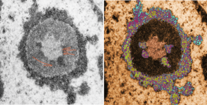 This image would be a fun cover submission… maybe someday i will make up a panel with three or four TEM look alikes. I know i have a valentine, and a ghost and a donut.
All grown up when—
Quoting a friend here who defined “growing up” being OK with
“not getting the recognition we hoped for
not completing the tasks we dreamed about
not seeing the world become a better place and
not receiving the unconditional love we deserved”
a rehash being grown up is:
“praising others
sharing knowledge
spreading fairness
loving everyone”
Fairness…!!! not just a human emotion
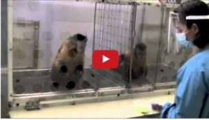
This is a lot of laughs (a TED Talk), but it speaks to every one of us… notice there is no gender mentioned for either participant, I think fairness transcends gender. It is worth a million admonitions from mom and dad, and from teachers, to help kids sort out fairness, and allow more to be given to someone else, but this demo packs a punch than all those verbal teachings put together cant override-because fairness is innate.
Granule sizes and spaces in the nucleolus
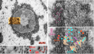
Epithelial cell in the lung of a baboon (probably a type II alveolar cell) showed a ring nucleolus with a single fibrillar center and concentric granular and dense fibrillar components. I took the time to measure (relative to a series of ribosomes measured in the adjacent cytoplasm – estimated at 27nm in diameter) the size of the granules in the dense fibrillar portion and in the granular portion of this nucleolus. The image on the left shows the whole nucleolus, the three divisions (there are probably more) of the nucleolus are prominent. From that image of the nucleolus, the orange area is enlarged twice on the right to highlight distance between granules of the dense fibrillar component and the granular component. The small redish box is what was used to estimate the size of those granules by measuring ribosomes therin (red dot) obtained from a portion of adjacent cytoplasm from the same image.
The diagonal (and rearranged and measured as horizontal) lines are those between granules in the granular portion (white) and also the dense fibrillar portion (purple). The spaces between the granules in the granular portion are marked out with a peach transparency and the blue circles represent the size of granules in that area.
Key: orange circles = size of the granules in the dense fibrillar portion at 30nm, with about 38nm distance between them; white lines and blue circles 21nm) represent the diameter of the granules in the granular portion and the distance between them (42nm). For comparison, the ribosome (red dot, lower center-left is 27 nm, and the distance between ribosomes is about 58nm. The apparently-coiled strings (pink) are something that occurred regularly, kind of an arcing gently oval beaded appearance. 13279_94-32_baboon_lung_PFP (perfluorophenanthrene).
Hysterical cartoon by someone out there… thank you
It is tough to get ditched: emerita
Boy, having been a woman in science for 50 years has been a trip. Not that women before me didn’t have it worse, cause they did. I could go on for several pages about how the University of Cincinnati, College of Medicine, Department of Anatomy (which of course has had many different chairs, names and focuses by now) really didn’t like women graduate students. In fact I was the only woman in a duration of 6 years to get a PhD there. I had male counterparts who were so inept that it took the help of many faculty mentors to get them through the program, I was not really supported in that way.
I remember my own advisor wasn’t particularly pleasant to me, and after I left to do a post doc made good on a grant with my “microenvironment for cartilage and bone development” idea. After my post doc in the Department of Environmental Health I went to Children’s Hospital (the Institute for Developmental Research, no longer an entity really, and the hospital also changed its name, and the hospital front has had two renovations over the original site which are neither better nor more beautiful than the old hospital. They did manage to save the stained glass windows and house them in a different location but I can remember what happened to the two large carved crosses atop the building, which came down with the wrecker ball). My immediate working salary compared to my similarly educated male counter part was an even half of what he was given. When I asked, I distinctly remember Leland Clark, Jr. saying, “take it or leave it”. Clark was another story, a hugely misogynistic egotist who would take info and publish it with out credit to colleagues, and could never do two things over again, everything was an invention (not good or bad, but terrible for a new career path in science). I watched him throw oranges, or pens down the hallway at the elevator door. I put up with his genius/insanity for 8 years, and what I got from Children’s was a letter after I took a position back in the Department of Environmental Health doing electron microscopy (Directors Dr. Suskind and Dr. Albert were both MDs and understood the power of electron microscopy…. after that, electron microscopy was not a convenient nor important tool… ha ha… so short sighted). When i left children’s I got a letter from Dr. Pratt…. “your services are no longer required”… that was it. No mention of the insanity from Clark that was tolerated.
The directors in Environmental Health have not been all that great… but for some reason the MDs sort of understood what was ultimately going to affect more lives in the near future than the later directors. The latter just don’t understand that we are still living in a world where some very inexpensive measures, and tools can impact many many children, workers, residents and change their lives so much. Instead they dove into the minutia, billions of bits of data, when teaching people to wash their kids hands frequently could have saved millions of IQ points. I am not saying minutia is not good, after all I have spent my life looking at objects in cells that are no more than 10nm… that is also minutia.
I did enjoy the old school faculty in the Department of Environmental Health on the 1980s to the early 2000s… then came the change in management (even from the top down) that took the “university” down the “business and profit” path, one of more repression (haha like the time George Leikauf, then director of Toxicology, came into my office and told me that I could not use WordPerfect, that I had to use Word…. i replied, George, are you telling me what kind of pencils I must use) than encouragement for individual thinking, and compartmentalization of the faculty into the secure (big brother tenured) and expendable (other adjunct, research and collaborative faculty) (not really based on merit), still heavily biased against women (Not to speak of trying to rear three children as a single parent — there were times I looked at one assistant director and thought to myself — all he has to do is pull on his pants in the morning then drive to work).
Two distinctly unfortunate events were both over ridiculous lies…. one from George Leikauf when in front of a site visit for the then Health Sciences Center, he implied that his electron micrographs were from the microscopy service that I ran at the time…. I told him in private afterwards that he could not do that, and besides the micrographs were not all that great and not mine. Haha… therin was the beginning of a struggle to get his recommendation for reappointments each year for the next decade…. his derogatory title for me was the Queen of Corel, because I used this program (which for some reason he didn’t approve of) for diagrams and illustrations for my work, the work of many colleagues and the progress reports and resubmissions of the grant.
Then there was the lie from one of the Acting Directors, Earnest Foulkes, when he stood in front of the faculty and said that I said that “so and so” was a great technician, which was totally untrue, had never had that conversation with him, and actually didn’t particularly like the work of that technician. I couldn’t figure out why he did that… but in private I confronted him… saying. you cannot get up and say things to the faculty like that which are totally untrue. Less than two hours later he stopped by my office and said “Marian, we can no longer pay for the service contract on the electron microscope” ha ha… give me a break. So I added that to the funding I had to dig up.
There were a few women who managed to survive here, but even the most well respected have been washed away with the waters of time. I guess in a sense, that is a huge waste of resources, and short sighted, but a matter of policy. UC, you have made changes, change yes, is a part of life (not all change is good, which is something that one hopes an institution of higher learning things about (but in this case, maybe not. Haha, like the hundreds of thousands spent on rebranding. oh my god, for what…! And not even that clever. Enough. I am deciding whether to contribute to the Foreign Department of Environmental Health or work on my own. Guess which… ha ha… I don’t know yet, maybe you can predict.
