Monthly Archives: August 2017

Perhaps just a little overzealous in praise of PFC-based blood substitutes

perfluorocarbon based blood substitutesHere is a quote from a chapter in “Blood Substitutes” (By Robert M. Winslow) and the chapter was written by Jean G. Riess, PhD and Marie Pierre Krafft, PhD. Please note that the location of the primary author is San Diego (home of an ailing Alliance Pharmaceutical Corp Oxygent PFC based blood substitute development) which (like an earlier post on an article by a researcher there – possibly the CEO) also read more like an advertisement extolling the virtues of a product they wanted promote than a scientific pronouncement outlining the effects, both good and bad, of PFC based blood substitutes. I quote one paragraph which is particularly off base, here ‘verbatim’.

Finally, PFCs are not metabolized, and no microorganism is known to feed on them – since Mother Nature did not exploit the PFC route, she did not develop the enzymes that would have been needed to recycle them. Pure PFCs have no effect on cell cultures either, other than the benefits that result from O2 or CO2 delivery. One can drink PFCs by the liter without side effects other than wet pants due to extreme spreadibility!

I take exception to several things here, firstly I have witnessed (with electron microscopy) in vitro responses in the reticuolendothelial cells to PFC:  1) and there are differences in PFC-induced responses, which vary with the PFC, and there are changes in the phagocytic cell population involved, including phagocytosis and the appearance of immune activation. I have seen macrophages that have engulfed enough emulsion particles so that they become half of the visible cytoplasm.  If this is “no effect” I will laugh — it certainly is an effect.  2) also, macrophages  produce visibly increased amounts of lysosomal enzymes that get piled into their phagolysosomes… as very dense caps.  Were that not enough, the last several posts on PFC and macrophages (in vivo) lysosomal inclusions have shown that a huge response in the multivesicular bodies/late endosomes/PFC inclusions to the PFC E2.

Image: cut and pasted and highlighted red cells, shadowed, and also wikipedia’s image of the molecule for perfluorodecalin, the most commonly used ingredient in many of the blood substitutes.  (BTW, none of the various concoctions really were viable substitutes).
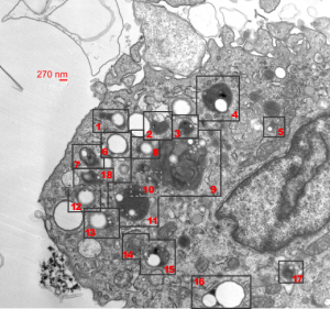
MVB/LE/PFC: multivesicular bodies, late endosomes, perfluorochemical inclusions

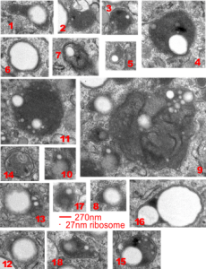
Alveolar macrophage from a mouse lung, the latter having been subjected to complete submersion in E2 perfluorochemical for three hours, and allowed to recover for 48 hours.

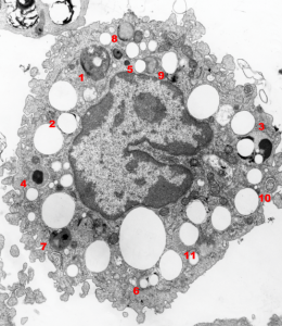
A single macrophage has almost all the PFC inclusions with high concentrations of enzymes, making them appear to have black “caps” in some cases, and in others, mostly enzymes, showing the tiny E2 particles and larger particles (does anyone know if this is Oswald ripening…. it seems as if the particles are doing two things, getting smaller and coalescing…??). The variety of multivesicular bodies here is quite amazing, with some having other structures included, some being filamentous, mostly being very very electron dense.  Each of the MVB/LE with PFC inclusions (i did not tag them all, it was too cluttered) have a bounding box indicated on the unretouched first image,  they are all enlarged and collected in a gallery below (numbered so you can find them).  Compare the variety here with three other posts on MVB/LE/PFC.

BOOM

Thus TWEETS the president: “Military solutions are now fully in place, locked and loaded, should North Korea act unwisely. Hopefully Kim Jong Un will find another path! ”   so sad to say but THESE ARE BOTH DUCKS OF THE SAME FEATHER.  Who says stuff like this except individuals who are just not in touch with reality, nor do they care about anything except their self esteem (which in both cases is below my shoe sole).  This is Lose-Lose…since we already know Kim Jong Un will be unwise, as will be Trump.

and did trump really tweet this in january… apparently so.

“North Korean Leader Kim Jong Un just stated that the “Nuclear Button is on his desk at all times.” Will someone from his depleted and food starved regime please inform him that I too have a Nuclear Button, but it is a much bigger & more powerful one than his, and my Button works!”   what a dumbnut.

Fruit loops

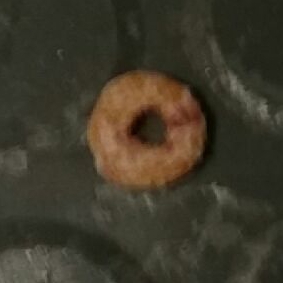
Aside from naming Howard “fruit loops” on the big bang theory, his official astronaut name, fruit loops can appear in strange places, to wit, on the back staircase of what has been called Kettering Laboratory, or the Department of Environmental Health, at the University of Cincinnati.  I have been tracking this little “fruit loop” for many months.  The progress it is making towards being swept up by the crew that cleans the back stairs is monumentally slow.

This reminds me of my best colleague ever, in electron microscopy research, fruit loopfrom about 1980 – the early 2000s, she and I monitored some dead bumble bee or wasp (or maybe is was a flying red roach) on the stairs in the oldest wing of the building (which is no longer standing due to the lack of foresight of the “visionaries”), at one point using a sharpie to circle it and date its appearance (this was after it had already been there for several years).
ONE more day and counting 8-9-17

I am taking bets on the fruit loop.  Maybe already about 3 months have passed…  I will keep you posted.  Ha ha.

fruit-loop is gone  8-10-17 (i think someone is monitoring my blog… ha ha)

MVB/LE/PFC in macrophages can be other than rounded

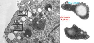
Multivesicular bodies in alveolar macrophages are generally roundish, not always perfectly round, but more round than elongated.  Here are two multivesicular bodies-late endosomes with inclusions which are presumed to be E2 left over from liquid breathing (MVB/LE/PFC) which have lots of protein enzyme content (way more in a volume ratio than E2) and have an elongated shape.  There is a large reaction to the E2 droplets in terms of enzyme production.  So the ratio of perfluorochemical to protein in the lysosomal structures might give a clue as to the characteristics of “export” or “offgassing” of these liquids from the body. The size of the smallest E2 particle in these micrographs is slightly larger in diameter than a ribosome (taken from the same micrograph and enlarged similarly (two insets at right are from the respective boxes top and bottom in the portion of the alveolar macrophage, and there are many MVB, different sizes and many E2 droplets with different amounts of enzyme.  I would guess that the smaller droplets are more enzyme packed than the larger droplets — equating to residence time within the macrophage cytoplasm???

COPI I

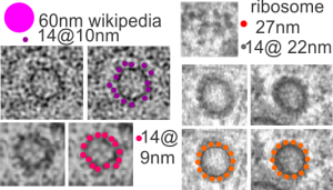
Found these two pretty nicely organized COPI I (probably) vesicles in an alveolar macrophage from a mouse which breathed E2 bubble oxygenated liquid for 3 hours and was allowed to recover for 48 hours.  This macrophage had particles (droplets) of E2 within but I cropped out the two vesicles which displayed pretty even symmetry and a close to equatorial sectioning.

These micrographs were all marked up with pen and lines and I rescanned it from the negative (1347_4840_2,500x, mouse, E2 breathed 3 hr, 48 hr recovery, electron micrograph).

I sought out some TEM images of COPI I vesicles and found a composite of in vitro vesicles on wikipedia here, and from images, this article on researchgate which offered up some info as well.  I think my images show the best symmetry, just by chance at about 14 densities per approximate equatorial circumference. Diagrams of COPI I vesicles don’t seem to offer an exact number of constitutive or incidental “blips” on the cytoplasmic side of the membrane, so this is just a guess. My dimensions were derived from adjacent ribosomes, taken at 27nm each as an average, Their calcularions?? but as an estimate perhaps those densities on the surface are 10-14nm in their rounded diameter.

MVB/LE/PFC bodies within alveolar macrophages of liquid-breathed mice

In this case the liquid which the mouse breathed for 3 hours and allowed to recover for 17 days was E2.  Thank goodness for good record keeping otherwise I would have forgotten what E2 stood for. Here is what the notations say (from back when Leland Clark Jr. was keeping tabs on all documents and publications.  E2 (made by Du Pont) O2 solubility ml/100ml at 25oC 55.7, vapor pressure 56mmHg at 37.5oC, viscosity 0.6 at 25oC, boiling point at 104.4oC, density gm/ml 1.656 at 25oC, surface tension 12.9 dynes/cm at 25oC.E2 liquid for breathing perfluorochemical molecular
There were a bunch of MVB/LE/PFC organelles in these macrophages that I began to look more closely at the parallel leaflet patterns that were showing up at higher magnification.

Altogether there were 29 mice that breathed E2, it much have been a satisfactory fluid or there would not be a dozen or more mice that survived from 5 days to 449 days after 3 hr of breathing E2.

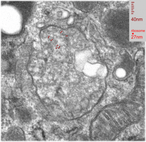
The electron micrograph below was one such body, I don’t think it could clearly be labeled a multivesicular body or a late endosome, but there are E2 droplets involved in its organization.  I don’t believe there is any outer nuclear membrane association, but there are membranes with different distances apart which are clearly organized (actually a parallel disorganization – if such a thing can (and it does visually here) exist).  So I sensed that the distance between the leaflets of these membranes was different within the MVB/LE/PFC thingie and in the membranes arranged just outside the MVB/LE/PFC structure and they were different. The latter look to be about separated at 22nm n=28, x=0.053+0.0014  (mm, on the micrograph translated to close to 22nm in actuality) , and I also measured couple of dozen distances between parallel pairs within the MVB/LE/PFC structure itself and it was closer to 28nm (n=31). When the long string of measurements were subjected to a t-test (remember this is an N of ONE macrophage (there will be others) so it is not really saying much) there was an highly significant (p=0.0001) difference between the mean and SEM of the two (color coded) groups. Pictures below show how the lines and data shaped up. White dotted line outlines the two areas sampled for leaflet separation.

Figure below that shows actual sites of measurement and place of measurement for each group.

liquid breathing electron microscopy mouse lung E2

liquid breathing electron microscopy mouse lung E2In another macrophage –another micrograph — another block same E2 treatment and duration, a similar type of inclusion is seen, but this one did not display the linear qualities of the MVB/LE/PFC as above.  There are two droplets of presumptive E2 in this image, they are middle right and upper left.  Whether the lucent area in this inclusion is E2 is anybody’s guess.

Mouse, liquid breathing E2: macrophage and lysosomes

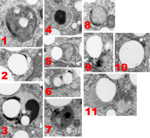
Here is a gallery of the types (big variation) of lysosomes that I found in a 1000A thick section of a single alveolar macrophage from a mouse that had breathed liquid E2 for 3 hours and then was allowed to recover for 17 days before euthanasia.

The only duplicate in style was found in the those MVB/LE/PFC (multivesicular body/late endosome/perfluorocarbon particle) that displayed a small very dense black cap, likely to be the distribution of the proteins.  Sometimes these contained one or two small droplets of E2 (6) or the densities were found at the margin of one large droplet of E2 (2) and even these have different morphologies. A less dense background and divided density contents of some MVB/LE/PFC bodies were NOT arranged with enzymes (proteins?) at the periphery only (3, 5, 7-9) and some polarity to the distribution of the darkest portion (more enzymes) of the MVB/LE/PFC. The lysosome 3 is quite interesting, with three definite PFC droplets and a very marked separation of a less electron dense background and dense compartment (presumably enzymes, maybe portions of some other proteins or even surfactant lipids.  Lipids would be a good guess since they are so darkly stained with the osmium tetroxide). The difference in the size of these droplets is interesting as well, since it seems that the smaller droplets (and maybe they shrink to not detectable with TEM) could be the ones which escape the cell and are offgassed in the lung.  I am just linking the complicated stuff on wikipedia about this, but you can be sure that the temperature difference between deep tissue and lung, and size differences in droplets makes a huge difference in vapor pressure…. someone else can figure this out).  Figure 5 below also shows the tiniest E2 droplets… actually interestingly also noted that the droplets are for the most part found on the periphery of the bounding cytoplasm (likely membrane bound, but perhaps the PFC just interfaces with adjacent proteins in a manner that looks membrane bound (that said for those droplets that do not LOOK to be inclusions in MVB/LE/PFC structures… but then, my guess is that they are all membrane bound having been taken in endocytically.

So the variation is huge, some, like image 1, look like there are lamellar body proteins resembling surfactant and lipids.

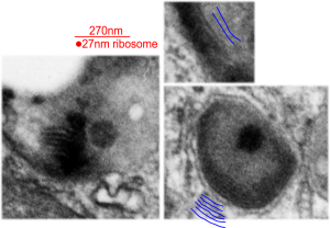
There are other MVB/LE/PFC structures which show a distinct lamellar nature with the distance from one dense line to the other is about about 32nm (see below). These are from 1, 9, and one not shown in the gallery above, but just cut off on the corner if image 1, but present in the whole alveolar macrophage at the bottom of this post.

Unbiased research? Publications? Pharma?

II was reading a pdf available online on perfluorochemical based blood substitutes, and also in general thinking about liquid breathing of perfluorocarbons. I decided to google the topic to see if any new research had surfaced and I found this 1994 article ” Stephen F. Flaim (1994) Pharmacokinetics and Side Effects of Perfluorocarbon-Based Blood Substitutes, Artificial Cells, Blood Substitutes, and Biotechnology, 22:4, 1043-1054, DOI: 10.3109/10731199409138801″ My guess is the journal is no longer active. But reading the pdf (yes i went to the literature cited section to see if there was any mention of the articles I had published on the topic, and while Leland C. Clark, Jr showed up, none of the morphological studies by MOI were cited) but it read like a sales pitch for the use of perfluorchemicals (which I personally never thought were that kind to the body). After reading about ten times phrases like “these effects are reversible” “no visible signs of interstitial edema or pulmonary lesions” and “no evidence of interstitial edema, necrosis or fibrosis” or “no organ damage is manifested” and “not regarded as significant side effects” I about gagged. What, is this the same stuff I did research on, found gobbled up to massive extent in all the reticuloendothelial cells, even in parenchymal cells, even tested as a stimulant to the immune system to reduce tumor graft growth. Is this the same stuff that I would not have put in my own veins or lungs under any circumstance? Ha ha…are we talking about the same thing here…. perfluorochemicals for liquid breathing, radio contrast, and blood substitutes.. yep. So I boldly googled the author… ha ha… there might have been some kind of “bias” in his article…. after all he had a vested interest in a company that was marketing the stuff. Alliance Pharmaceutical Corp……. not good… and its really good that they are defunct.

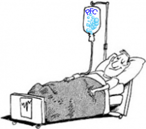
Stephen Flaim: From scientist to prolific entrepreneurblood substitutes modified cartoon

I also found this amazing blog which anyone interested in the topic should read. And felt compelled to modify their cartoon and when requesting permission the blog was 404… so who knows whose it is.

4 things learned– watch out for scientific bias, keep up your blog posts, be careful about what you let the MDs do, big pharma rarely helps.


E2 liquid breathing: no joke

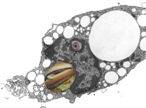
I really am fond of being silly, particularly when images like this hungry alveolar macrophage with eye and open mouth show up in my files.  I probably took this image for my archives on liquid breathing way back in the late 1970s, never seeing the humor in the anatomy.  But this morning when I was trying to figure out how to post this image, i just laughed out loud to see the perfect eye and open mouth and could not resist adding a “big mack” and “eyeball” with photoshop.  The original, actual serious micrograph is below the cartoon.

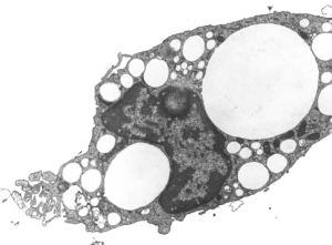
So here is an alveolar macrohage, in a pretty good looking alveolus, from a mouse which was dunked in E2 for three hours (bubble oxygenated) and survived and was allowed to recover for 17 days.  The pattern for E2 clearance from the lung is a little unique in that the droplets of perfluorocarbon are neatly coalescing into bigger and bigger droplets in some instances, but in others are seen as small droplets within, what i used to call “black cap lysosomes” which were apparently lysosomes with dense collections of enzymes, pushed over to one side of the organelle.  Thus is was assumed the perfluorocarbon droplets became part of the lysosomal pathways. The E2 droplets here are white, I did not pseudocolor them as in the previous post, I did remove three pieces of junk using the stamp tool in photoshop.  In this case the “eye” or “eyeball” seen for real in the bottom images is likely a tangential section of a medium size E2 droplet just pressing in on the nucleus making a symemtrical indentation. It does not have the character of a nucleolus, which the other option would be. The mouth of the cartoon was a reasonably large droplet of E2 (before i filled it in with big mack) probably on the order of 3-4 microns in the observed diameter. The tiniest droplets of E2 can be seen here and there in the cytoplasm, some of them even less than 0.5 microns in diameter. Mitochondria in these macrophages do not seem to be stressed by the presence of E2 droplets (more or less confirming their alleged inertness) and the amount of nuclear condensed chromatin would also suggest that the macrophages are not activated by E2 since there seems not to be any ramped up RNA, ribosomal, synthesis in the nucleus, or protein synthesis in the cytoplasm, which would suggest an increased synthetic activity related to an immune response to the presence of E2.

cartoon big mac mouth macrophage transmission electron microscopyAnd here is the scientifically sound, non cartoon, image.

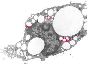
E2 within an alveolar macrophage electron microscopyHere is an image with four droplet or droplet groups which show the lysosomal enzymes highlighted in red. These observations relate to perfluorochemicals and fluorochemicals in general as the ability of the body to rid itself of them (whether through outgassing through the lungs reportedly in some relations to the vapor pressure and lipophilicity, and molecular weight. Other elimination is not reported to exist since perfluorochemicals are supposedly not metabolized, so metabolic degradation may not play a role here. I appears that how well the lysosomes “re-emulsify” the droplets is also relevant. Case in point for FC47…  it really never was successfully re-emulsified, as opposed to perfluorodecalin which made nice progress in the process of forming tiny droplets in pretty uniform size during elimination. Here you can see the even roundness and lack of droplet substructure and marginalization of the lysosomal enzymes — all important characteristics of the lesser-evil perfluorochemicals.

E2 within an alveolar macrophage electron microscopy lysosomes pseudocolored