Monthly Archives: April 2017

Great stuff, stuffed everywhere

This is “what not to do” with an old can of Great Stuff, you know that expanding foam filler for cracks and places that one doesn’t know how to seal.  It is without a doubt, a one-time-use fix-it thing, as i had two left over cans from other long ago projects.  I wanted to seal a space that had widened between the stucco and my tiny back porch roof and googled all different methods and the expanding filler turned out to be a reasonable solution, perhaps.

I had saved the little plastic squirt thing, all cleaned and nice, hoping that when I needed it again it would work, but not really. It was extruding from that tube so sloooowwwwly that i was getting exhausted trying to make it work. Then (as happens very often in cincinnati) it started to rain. I knew the moment i stopped extruding that stuff that it was “game over”, and of course I was right.  So i hunted around the house hoping to find a substitute through which to press the remaining great stuff into the space…. and i found another half used can.  This can would not let me press the rubber gasket to get anything out at all, so i just pressed it as hard as i could against a rock (outside), and woila, out it came, so now it some coming out without restraint…  at that moment i tried to capture some and press it into the space, what a mess, and great stuff just kept coming out and i had no way to stop it…  i did press enough into the crack to fill it, and tossed the whole can into the garbage against some foam from a seat that the dogs had destroyed…. I wanted to save my trash can  (LOL)… and here is what you get when “great stuff” empties itself into the garbage over the next hour +…  By the way, the crack sealed just fine…. I re-primed the flashing, but it took several days to get the gunk off my hands.  Not the most efficient repair job in the world, in fact funny at worst, hysterical really.

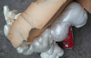
So next step was to google how many accidents with great stuff have been registered online… and of course I could have guessed that it is a medium for sculptors…. my next project will be a great stuff christmas tree.

..yes, one more cover submission illustration — I just love these granules

..yes, one more cover submission illustration — I just love these granules, and I think this might be my favorite yet. Colored all the granules in (using photoshop) overlying their own original images from the basic research on this putative SP-A granule, and arranged them in a highly ordered format (haha) which is certainly not really my normal MO, since I am kind of a random thinker.  But this is nice…in my opinion, and also a little artsie-craftsie for a scientist.. but thats what covers for scientific journals are for.

…and yet another cover submission for the manuscript on SP-A granule and electron microscopy

…and yet another cover submission for the manuscript on SP-A granule and electron microscopy.  This one is a little more dramatic than yesterdays post from the bottom right part of the image.  I like it better. It shows the quintessential granule, in black and white at the top, color at the bottom in a vertically mirrored image, directional ribosome attachment, electron-lucent areas under the non-oligomerized protein part, distinct periodicity to the dense bands (three complete “periods” in the lengthwise portion of this granule) and also the periodicity to the dense layers (3-4 dots/100nm) and even some of the faint periodicity found in the central lighter layers. In this electron micrograph one also sees part of the nucleus (bottom), a couple of nuclear pores, and some perinuclear space (but no granule formation in the perinuclear space is seen in this image, but it is found there frequently) and on the top, portions of three mitochondria and one adjacent to the lucent area of the granule. There are also other profiles of RER, coming and going which at some point might connect up to layered putative SP-A protein granule structures.

Four cover submissions for the SP-A manuscript: love the colored electron micrographs of granules

Four cover submissions for the SP-A manuscript: love the colored electron micrographs of granules. I am submitting this to Anatomical Record. They request a cover submission at the same time as initial manuscript submission, apparently, at least those are the instructions.  Typically I think the manuscript has to be accepted before cover submissions are made.  Oh well, I am just hoping for the best.  Preview here.

And there you have it!

Biggest bomb ever, dropped by the the biggest idiot ego ever. This is not my kind of president, and not my kind of American. So sad, and it certainly does not mesh with how I would help any war torn nation.  Maybe some of the security $$ from the Florida white house should be used for immigration, and safe passage and peace negotiations.

I do believe there must be some underlying bullying that occurred in this mans life that makes him behave like a bully himself.  Lord help all those Americans who don’t like the track this administration is taking us.


3 diagrams as putative fits for alveolar type II cell granule

So tired of looking at this structure and trying to brainstorm about how things fit together and whether SP-A is a good protein fit for this very regular and very nice molecular alignment within the RER of alveolar type II cells of several species.

A. Single period (RER membrane on top and bottom) from ferret show outer dense layers and less dense central layer. B is also a single period of a granule from guinea pig which is not bounded by RER but by another period above and below. Less electron-density accompanies of outer dense layers occurs when adjacent periods are present. C is an end on view of a cylindrical granule from guinea pig, almost 200 nm in diameter which could be twice the single period width. The inner dense central layer would be compressed into the central density. The RER membranes are seen on the upper and left on the outer edges, and periodicities for both the less dense center layers and dense layers appear as concentric rings. D, is the same image as C, with a cylindrical model of an end-on view of the possible arrangement of SP-A molecules which could account for this structure. The number of densities in the concentric layers was used to calculate how many molecules to use for the model. Vertical lines (shown in Fig. 5 as well) became radial spokes. Bars = 100 nm. E. On rare occasions, and only in guinea pig, round and dense fuzzy-ball like structures were seen squeezed between the typical 100 nm periods. Box in E was enlarged in F to show the relative sizes of the round densities. A diagram of a potential SP-A fuzzy ball, approximately 30-40 nm in diameter approximates measurements of the spherical densities. Grey circle is ribosome size for comparison. Bars = 100 nm

Figuring out the truth

Feeling like our inability to tell fake news from real news:

If the news is reduced to “he said she said” maybe we are less likely to think we can figure out the truth, maybe then we just stop trying to figure out the truth.

Electron micrographs of golgi and golgi stress

Cover submission from the early 2000s for a published manuscript by Shull Miller and Prasad where golgi, golgi ion transport, and golgi membranes (yep, i am not capitalizing the word Golgi any more, with all due respect to Dr. Golgi, this is no longer about him, it is about the organelle and he has had plenty of fame already).

This cover submission was about flattened golgi, cis and trans faces nicely curved, as opposed to dilation and collection of protein contents.  The cover was never executed, but it is a fun graphic, none-the-less.

Photoshop was used to change color gradients of different electron-densities of the membranes and contents. Cytoplasm here primarily, only a portion of other organelles.

There is an obvious texture to the area associated with the dilated ER, how fun it will be (maybe it will happen) to return to those micrographs and hunt for a patterning in the protein aggregates.